
Musculoskeletal Quantitative Imaging Research (MQIR)
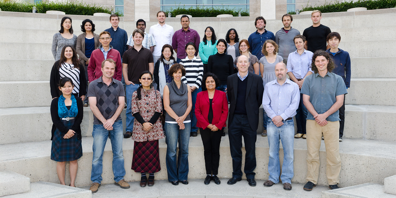
The Musculoskeletal Quantitative Imaging Research (MQIR) group consists of faculty members, postdoctoral fellows, research staff, and medical and graduate students who pursue their passion for teaching and research in quantitative tissue characterization focused on the musculoskeletal system.
Sharmila Majumdar, PhD, and Thomas Link, MD, PhD, serve as Director and Clinical Director respectively and lead MQIR’s aims to integrate research and build collaborations between basic scientists, clinical scientists, and physicians, establishing a strong resource for musculoskeletal imaging research at UCSF. Recognizing that collaboration is the key to advancement in today's research climate, MQIR builds partnerships within the Department of Radiology and Biomedical Imaging and with the Departments of Orthopaedic Surgery and Medicine at UCSF and Bioengineering at UC Berkeley.
Research
Musculoskeletal Quantitative Imaging Research Directions
- Identify biomarkers for bone, cartilage, and disc degeneration and diseases that impact the musculoskeletal system – osteoporosis, osteoarthritis, diabetes, and HIV.
- Develop new imaging techniques for bone marrow adiposity with MR spectroscopy and cortical macro-porosity using high resolution peripheral QCT to better understand fractures.
- Detect early joint degeneration and explore altered joint biomechanics and cartilage degeneration from sports injuries.
- Advance technical development of magnetic resonance imaging (MR), high-resolution peripheral quantitative CT (HR-pQCT), positron emission tomography PET/CT and PET/MR imaging, computer vision, image processing, and artificial intelligence (AI) to better understand musculoskeletal health and study risk factors for aging and disease.
Musculoskeletal Quantitative Imaging Research Labs
People
Directors

Radiologists
Imaging Scientists


Contact Us
MQIR Contact
Mission Bay Campus
1700 4th St., Suite 203
San Francisco, CA 94158
Ph: (415) 514-9655
Fax: (415) 514-9656
China Basin Campus
185 Berry Street, Suite 350
San Francisco, CA 94107
Ph: (415) 353-9401
Fx: (415) 353-9423