


Message from the Chair
Wellbeing & Professional Climate

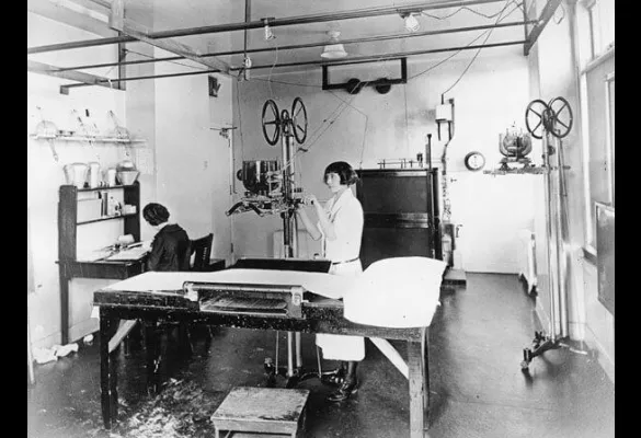
UCSF acknowledged radiology's vital role in patient care over 100 years ago by opening a dedicated x-ray facility and making sure all medical students were instructed in radiology. (Circa 1920)
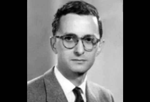
UC further committed to radiology as essential to healthcare by appointing Robert Spencer Stone, MD, as its first full-time faculty member. Dr. Stone served UCSF from 1928-1964.
Dr. Earl R. Miller served UCSF from 1943-1974.
(l-r) Drs. Hedvig Hricak, MD, PhD, Alexander R. Margulis, MD (Chair from 1963-1989), David Norman, MD and Charles Higgins, MD.
Dr. Thomas Hans Newton served UCSF from 1959-2009.
Having proved the usefulness of CT scanning, the UCSF Department of Radiology funded a small R&D operation, tasking engineers and physicists with developing nuclear magnetic resonance (later known as MRI) as a viable imaging instrument for soft tissues.
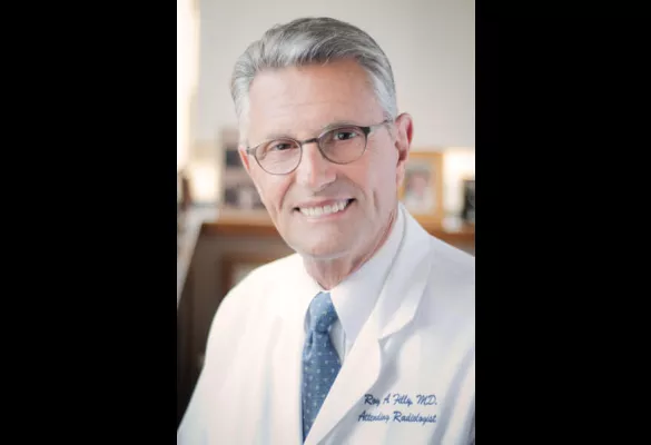
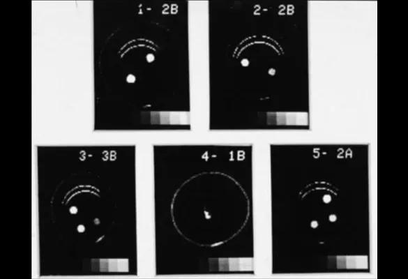
The first fetal surgery was performed at UCSF by surgeon Michael Harrison, MD, and radiologist Roy Filly, MD. In this surgery, an ultrasound-guided surgical opening to the fetus’s bladder (vesicostomy) was made. The UCSF Fetal Treatment Center, the first such center in the US, was co-founded by Drs. Filly and Harrison in the early 1980s and is now entering its fourth decade.
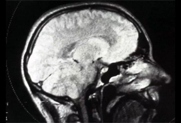
Radiology made the leap from imaging bones and hard structures to visualizing soft tissue with the introduction of the MRI. Much early research and development for this tool was conducted at UCSF.
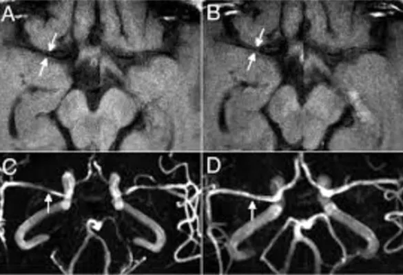
UCSF researchers developed methods of using MR to image vessel walls inside the skull, enabling more accurate diagnosis for stroke prevention and treatment.
Dr. Nola Hylton performed the first breast MRI at UCSF. Breast MRI is the most sensitive imaging test available to detect breast cancer.
UCSF research scientists led by Bruce Hasegawa, PhD, combine SPECT functional imaging with CT anatomical imaging to produce the first dual-modality imaging system, SPECT/CT.
Christopher Dowd, MD, and other UCSF physicians developed image-guided retrieval of stroke-causing blood clots.
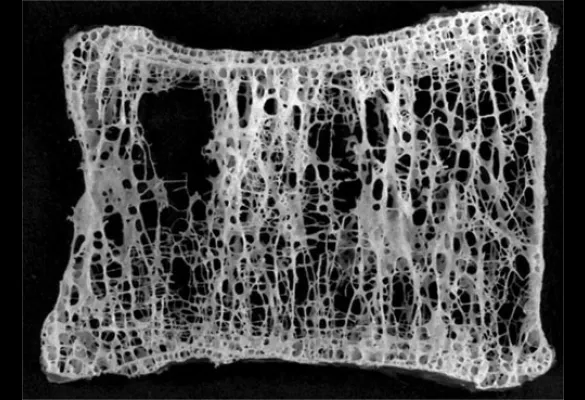
Thomas Lang, PhD, and colleagues from UCSF and Baylor School of Medicine began work with NASA on bone loss in astronauts, research now applied to earthbound bones.
The NIH funded UCSF's purchase of the country's first MR-guided focused ultrasound surgery system for surgeries using soundwaves to burn tumors.
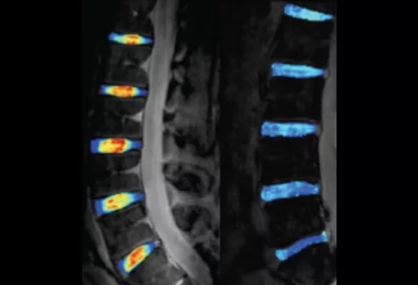
Studies began at UCSF on the use of T1ρ quantitative MR imaging, allowing the detection of cartilage injuries not visible with conventional MRI before tissue loss begins. Today, the technique is broadly used to create more treatment options and hasten recovery.
For the first time in the US, full-body, simultaneous time-of-flight PET/MRI became available at UCSF. This state-of-the-art dual imaging technology reduces radiation, enhances image quality, and is safer and more convenient for patients.
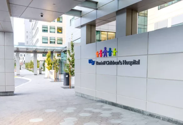
The new UCSF Mission Bay hospitals, comprised of UCSF Benioff Children’s Hospital, UCSF Betty Irene Moore Women’s Hospital, and UCSF Bakar Cancer Hospital, opened in February 2015. The Department of Radiology and Biomedical Imaging opened UCSF Imaging Center at Montgomery Street, a small, personalized site featuring screening mammography, bone densitometry and ultrasound.
In 2017, Ronald Arenson, MD retired as department chair, after 25 years. His chairmanship has marked a period of growth and innovation including opening of the Mission Bay campus and industry-wide deployment of a picture archive and communications system (PACS).
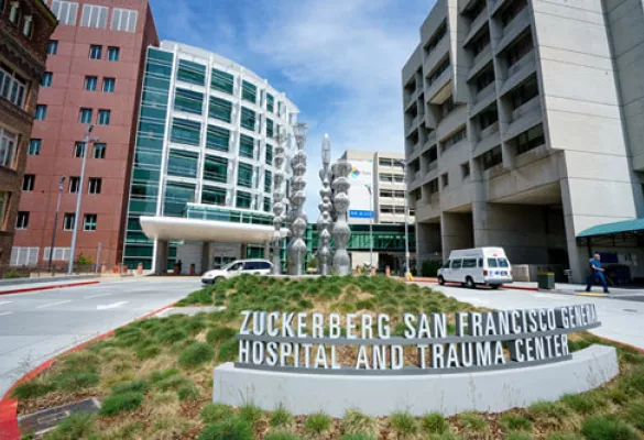
The new Zuckerberg San Francisco General Hospital and Trauma Center (ZSFG) building officially opened its doors in 2017. The new design was created to facilitate excellence in patient care with three floors incorporating Radiology.
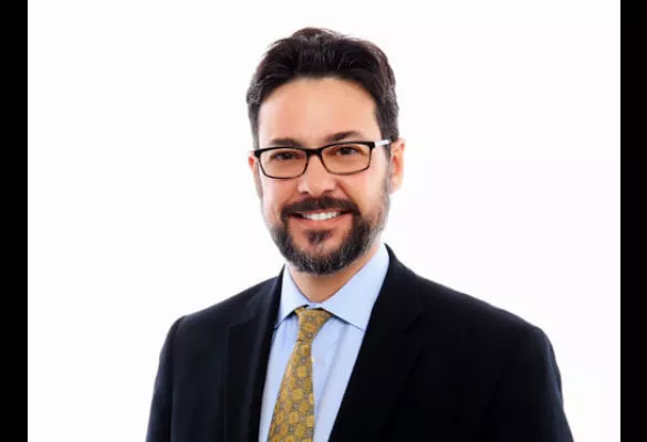
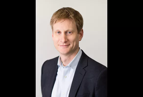
Christopher P. Hess, MD, PhD, was appointed as Chair of the Department of Radiology & Biomedical Imaging and assumed the chairmanship on January 1, 2018. In this role, Dr. Hess became the Alexander Margulis Distinguished Professor of Radiology.
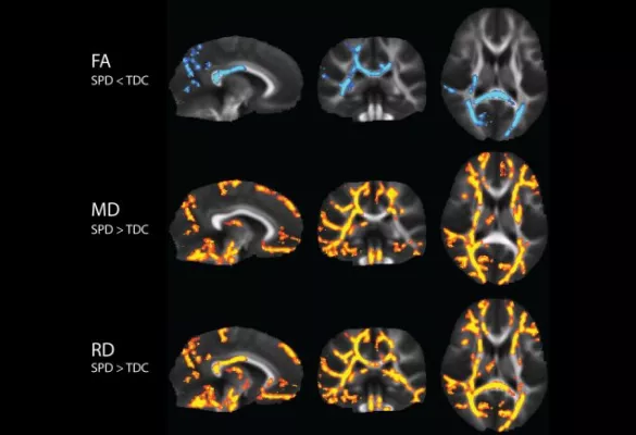
Pratik Mukherjee, MD, PhD, and his team’s groundbreaking work on Sensory Processing Disorder (SPD) published in Frontiers in Neuroanatomy, was the biggest imagng study ever done in children with the condition. His research promted a broad acknowledgement of SPD as a separate disorder from autism.
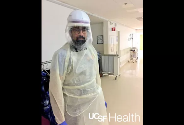
Protecting the health and safety of our patients and our teams is of the utmost importance with the emergence of COVID-19. Dr. K. Pallav Kolli has played a key role in COVID-19 preparedness work at UCSF Health and in the Department of Radiology and Biomedical Imaging where he is the Associate Chair for Quality and Safety.
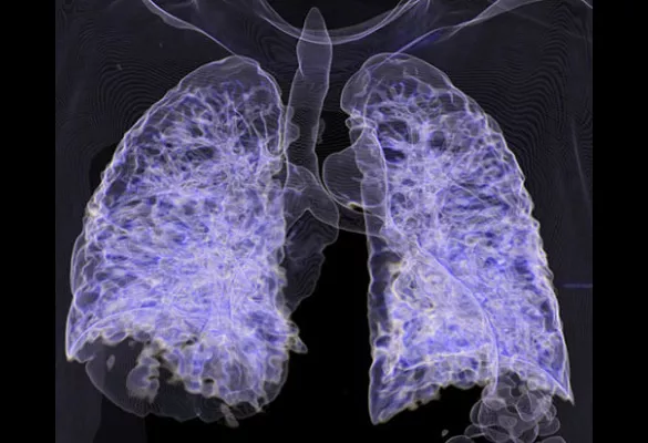
3-D CT COVID-19 in the lungs.
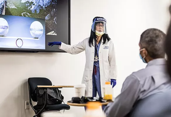
The resident and fellow orientation on July 1, 2020, a few months into the COVID-19 pandemic, required new safety protocols. Here, Dr. Soonmee Cha presents to new trainees on their first day in the department.
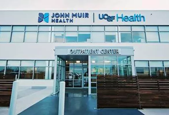
UCSF Radiology expands imaging at Berkeley Outpatient Center (BOPC) October 2020.
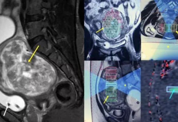
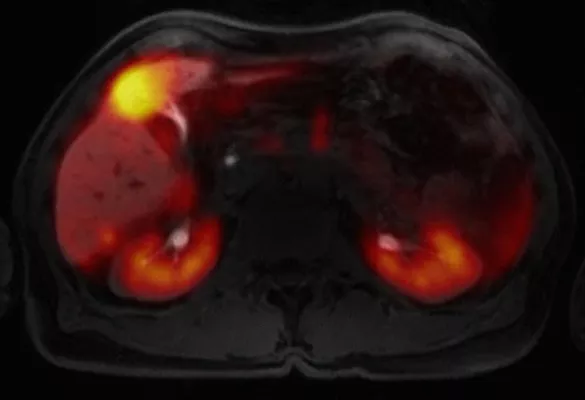
Dr. Thomas Hope leads breakthrough PSMA PET imaging for prostate cancer treatment, September 2020
In July 2021, the Alzheimer’s Association awarded the Henry Wisniewski, MD, PhD Lifetime Achievement Award to Michael W. Weiner, MD for his outstanding achievements in Alzheimer’s Disease research.