
Imaging Research for Neurodevelopment
Led by Dr. Duan Xu, the Imaging Research for Neurodevelopment Lab at UCSF focuses on early detection of abnormal development in newborns. To achieve this goal, the researchers in Dr. Xu's lab develop dedicated MRI hardware, pulse sequences, and postprocessing techniques. These methods are then applied to the Premri, Bamri, and Cardiac cohorts to further elucidate neurodevelopment of the neonatal brain.
UCSF Radiology: Baby Brain Research Group
Science
BAMRI
This study explores hypoxic-ischemic brain injury in term new born infants. Hypoxic Ischemia Encephalopathy is a type of injury caused by lack of oxygen and/or blood supply which can occur before or during birth.
PREMRI
Currently there is no way to predict developmental outcomes in babies that suffer brain injury as a result of prematurity. The goal of this study is to see if we can identify a relationship between the findings on a baby's MRI scan and his or her long term neurological and developmental outcome.
CARDIAC
The focus of this study is to determine if the aforementioned techniques can help detect brain injury in babies undergoing surgery for congenital heart disease.
C13
Our group is working with the Hyperpolarized Technology Resource Center to employ advanced C13 spectroscopy to study the metabolism of brain injury in rodents.
Engineering
Compact 7T
Imaging for Neurodevelopment is partnering with GE and the Mayo Clinic to develop and test the next big development in advanced brain imaging: Lightweight, Compact, Low-Cryogen Head-Only 7T MRI. Delivering powerful imaging capability in a small footprint, this technology represents a tremendous step towards bringing high-field imaging to more clinical and research settings.
Machine Learning
Can machine learning approaches improve how we gather, learn and predict with our infant MRI data? Our group is working to implement the latest advances in machine learning powered image reconstruction, processing and analysis.
Research
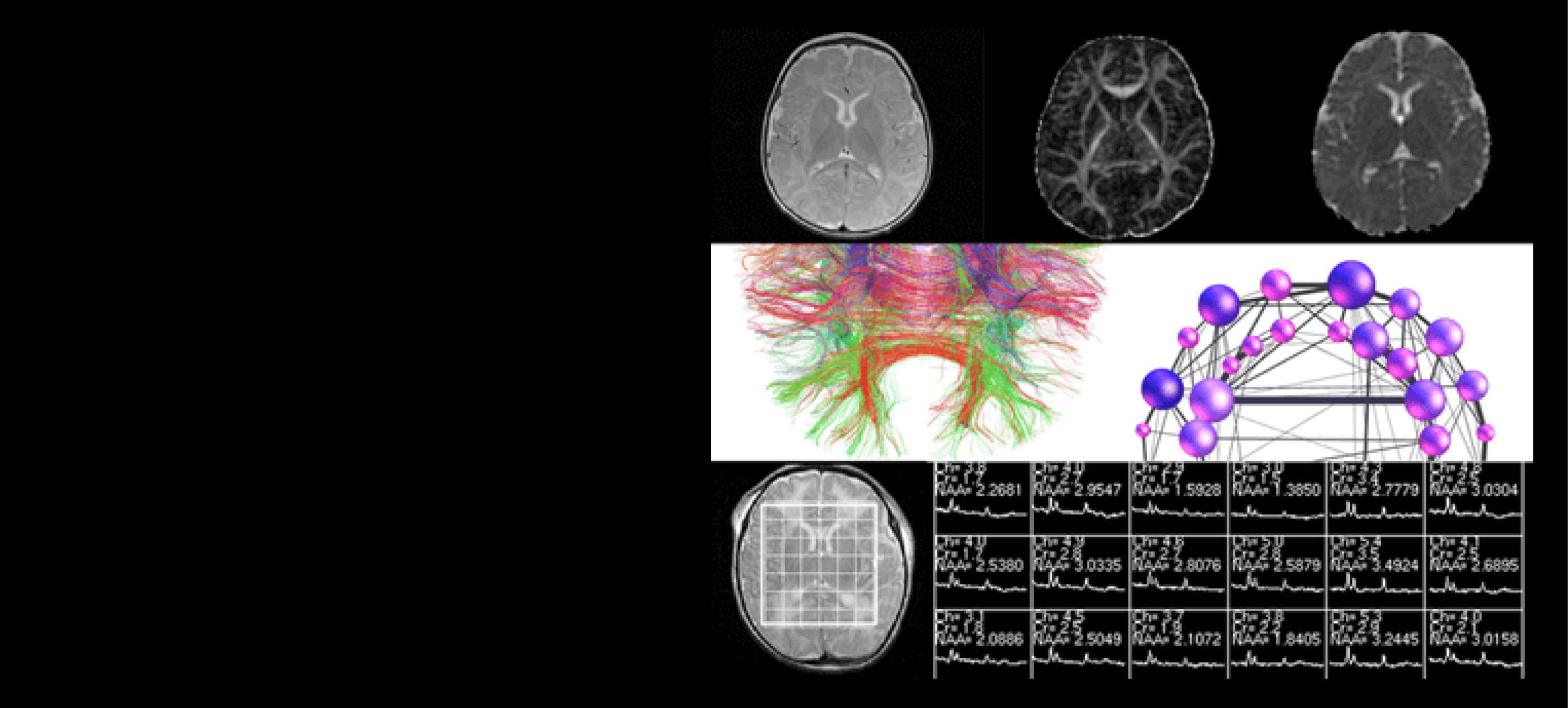
The goal of the project is to apply functional MRI technique based on BOLD mechanism to detect brain networks in babies. In order to do so, we detect temporal correlation between BOLD…
This project aims to modify and apply existing ASL techniques to study neonatal cerebral perfusion to better characterize infants with Hypoxic Ischemic Encephalopathy.
Studies have illustrated prognostic value in determining metabolite ratios between Lactate (Lac), N-acetyl aspartate (NAA), and Choline (CHO) from newborn infants.
The goal of this project is to establish a framework for assessing structural connectivity in the newborn brain at any stage of development, starting with premature neonates, and to…
Publications


Alumni


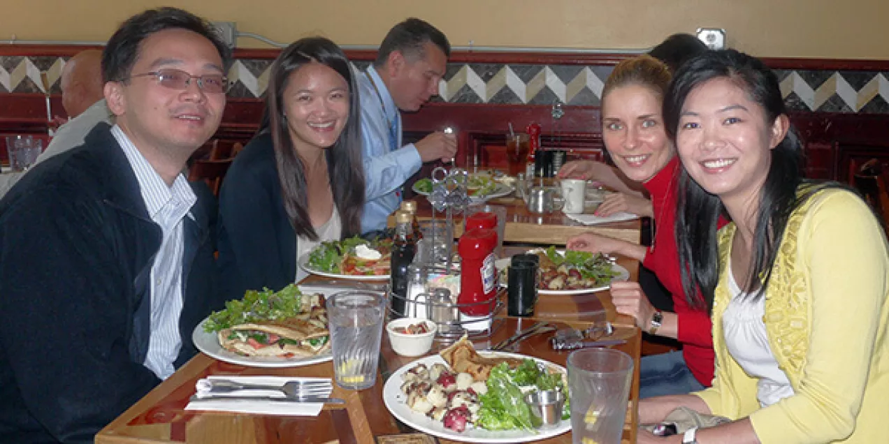
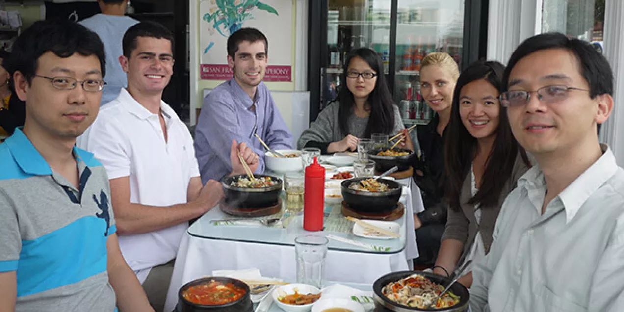
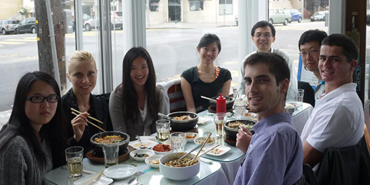
Lab Life & Team Moments
A collection of photos capturing our lab members over the years — from group meetings and collaborative brainstorming sessions to casual lunches and team gatherings. These moments reflect the camaraderie, diversity, and dedication that drive our research community.

Contact Us
Imaging Research for Neurodevelopment
Parnassus Campus
1 Irving Street, Suite AC-106B
San Francisco, CA 94143
Mission Bay Campus
1700 4th Street
Byers Hall Suite 102
UCSF Box 2512
San Francisco, CA 94158