Pediatric Radiology
UCSF Pediatric Radiology provides specialized imaging for infants, children and pregnant women. Recognized as the one of the nation's top ranked hospital, UCSF Benioff Children’s Hospital, we offer a unique pediatric focused environment that is safe and pleasant for patients and their families. The Pediatric Radiology division is dedicated to the health of children, conducting research, and training the next generation of radiologists.

Why choose Pediatric Radiology at UCSF
- Accurate and timely image interpretation and diagnosis
- Comprehensive services for all diseases
- World-renowned subspecialist in pediatric radiology
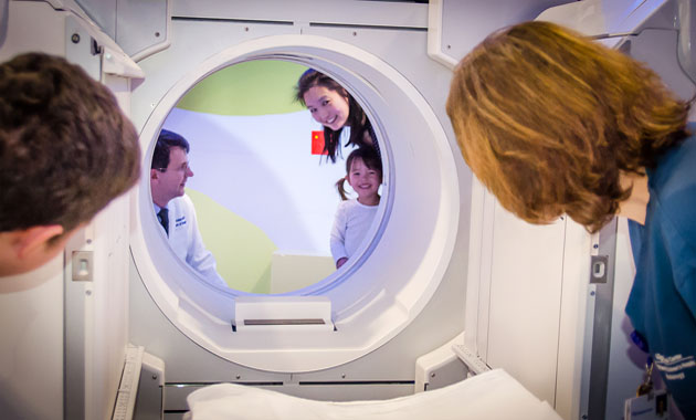
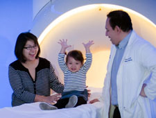
- Child–centered imaging, including child-oriented imaging suites, and partnership with UCSF Child-Life Services
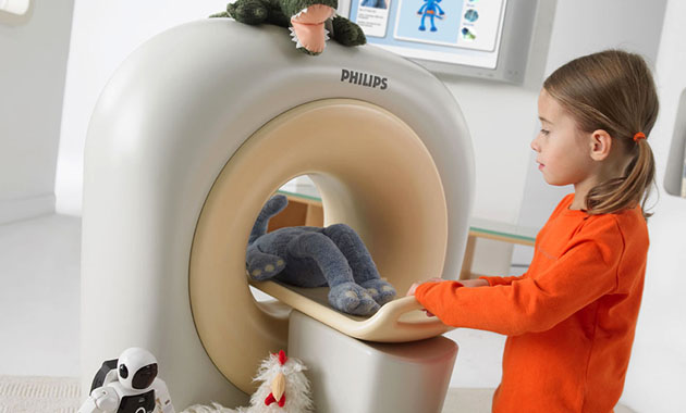
- Using the latest technology to reassure children and their parents
- Minimal use of anesthesia, sedation, and radiation
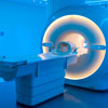
- Young patients can watch movies during their MRI procedure, minimizing need for anesthesia
- Multi-disciplinary teams with a wealth of experience and training in pediatric medicine
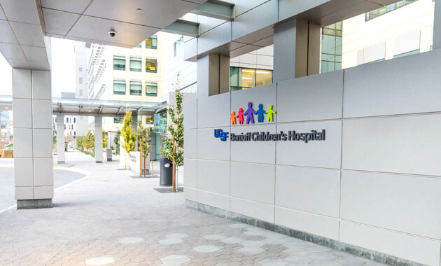
- Located at UCSF Benioff Children’s Hospital at Mission Bay and UCSF Benioff Children’s Hospital at Oakland, ranked among the top children's hospitals in US
- UCSF Pediatric Radiology is also uniquely designed to help mothers and transition their babies from fetal to neonatal life
Who we serve
- Pediatric patients and their families
Conditions we address
- All conditions affecting children
Pediatric Imaging Services
- 3D MRI, 3D CT, Augmented Reality and 3D reproductions using 3D printing technology
- Advanced high-field 3 Tesla MRI of the chest, body, and extremities
- Cardiac Imaging
- Fetal MRI
- Pediatric ultrasound – high resolution ultrasound, including advanced applications
- Pediatric focused X-ray, CT and MRI of the bones and joints
- Pediatric MRI of bowel, abdominal, and pelvic conditions– including advanced applications
- Pediatric fluoroscopy – including low radiation dose techniques
- Pediatric CT – including advanced applications and low radiation dose techniques
- Pediatric Orthopedic Imaging
- Pediatric Spectroscopy
- MR imaging with blood pool and bile duct contrast agents
- Magnetic Resonance Cholangiopancreatography (MRCP)
- MR Enterography
- Nuclear Medicine
Pediatric Imaging Safety
- American Academy of Pediatrics X-ray Shields No Longer Recommended
- NCRP Recommendations for Ending Shielding During Pelvic and Abdominal Radiography
Who we partner with
- Pediatric patients and their families
- Researchers from our own and other institutions
- Donors and other visionaries committed to improving the lives of others
- Referring colleagues, including general practitioners, peditricians, neonatal experts, and genetic counselors
- Subspecialists in cardiology, gastroenterology, interventional radiology, neonatology, nephrology, neuroradiology, nuclear medicine, oncology, orthopedics, pediatric surgery, primary care/pediatrics, rheumatology, and urology
Who we are
- Child life services and therapists
- Faculty members
- Medical and graduate students
- Pediatric administrative staff
- Pediatric anesthesiologists
- Pediatric nurses
- Pediatric radiologists
- Pediatric radiology fellows
- Pediatric technologists
- Postdoctoral fellows
Pediatric Radiology Faculty


Related Content
Pediatric Radiology Locations
UCSF Benioff Children's Hosptial
at Mission Bay
1975 Fourth Street
1st Floor Room C1408
San Francisco, CA 94158
Ph: (415) 476-1562
Hrs: Mon-Fri, 6:30am-9pm
Saturday hours available for some services
Emergency Room: 24/7
UCSF Benioff Children's Hosptial
at Oakland
747 52nd Street
Oakland, CA 94609
Ph: (510) 428-3410
Pediatric patients are our top priority
Refer a Patient
Radiology Scheduling
Ph: (415) 353-3900
Fax: (415) 353-7299
MyChart
Billing & Insurance
(415) 514-8888