
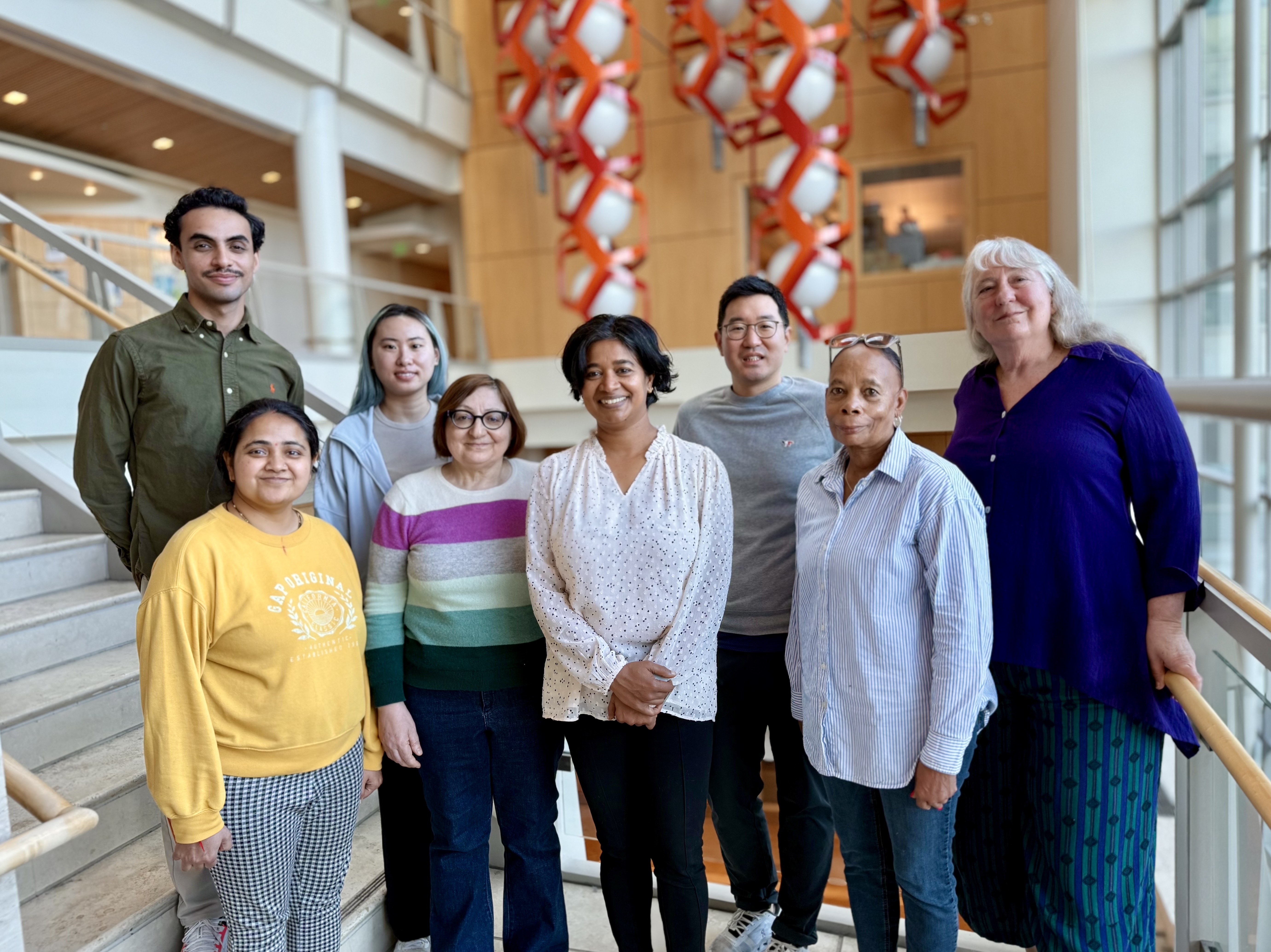
Translational Metabolic Imaging Lab
Guided by Drs. Renuka Sriram, John Kurhanewicz, and Donna Peehl, our team investigates how altered metabolism drives disease and how it can be visualized non-invasively to advance diagnosis and therapy. Our lab bridges molecular, cellular, and in vivo imaging modalities to develop metabolic biomarkers that can translate from preclinical models into clinical practice.
Research
The Translational Metabolic Imaging Lab focuses on developing advanced imaging techniques to better understand metabolism in health and disease. By combining imaging, biology, and clinical research, the lab aims to improve how diseases like cancer are detected, monitored, and treated through more precise and personalized approaches.

Our Research
We exploit metabolic dysregulation in cancer to identify imaging biomarkers of disease presence, progression, and therapeutic response.
Co-Clinical Imaging Research Program
To overcome translational barriers, we develop co-clinical imaging research that encourage consensus on how imaging methods are optimized to improve the quality of imaging results for co-clinical trials.

Publications
Opportunities
Rotation opportunities are available to get a feel for the multiple facets of a translational metabolic imaging lab.
People


- Joao Piraquive Agudelo
- Emilie Decavel-Bueff
- Kayvan Keshari
- Bertram Koelsch
- Vickie Zhang
- Avantika Sinha
- Jinny Sun
- Jessie Lee
- Rahwa Imam
- Justin De Los Santos
- Jeff Hsiao
- Fayyaz Ahamed
Additional Resources
Contact Us
Translational Metabolic Imaging Lab
Renuka Sriram, PhD
Associate Professor
[email protected]
Donna Peehl, PhD
Professor
[email protected]
John Kurhanewicz, PhD
Professor Emeritus
[email protected]