Neuroendovascular Surgery
Neuroendovascular Surgery (NES), also known as Neurointerventional Radiology, or Cerebrovascular Surgery, treats vascular diseases of the central nervous system.
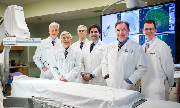
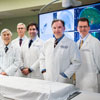
Our NES team is comprised of world-renowned physicians including experts from Radiology & Biomedical Imaging and Neurosurgery. Our team members work collaboratively to offer our patients state-of-the-art treatment to address a variety of conditions including brain tumors, cerebral aneurysms and other complex disorders of the brain and spinal cord.

Why choose Neuroendovascular Surgery at UCSF
- Endovascular techniques that lower the risk to patients, shorten hospital stays, and hasten patient recovery
- Accurate image interpretation and diagnosis
- Patient–centered imaging
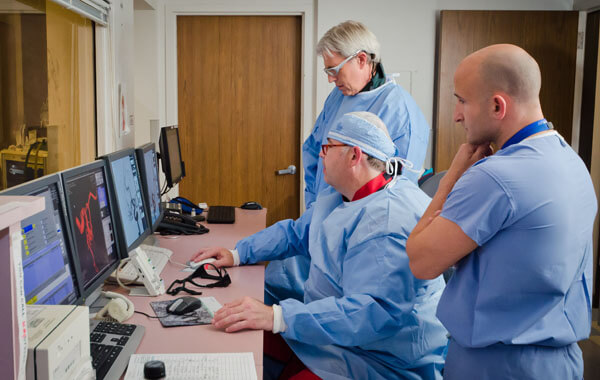
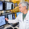
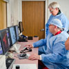
- UCSF angiogram suites are state-of-the-art with the ability to produce highest quality images to evaluate and treat cerebrovascular disorders
- Advanced procedures regularly performed at UCSF
- Ongoing development of new diagnostic and endovascular treatments for disorders involving the blood vessels of the brain, head, neck, and spine
Conditions we address
- Arteriovenous Malformations (AVMs)
- Birthmarks and Vascular Malformations
- Brain Aneurysms: Ruptured and Unruptured
- Brain Tumors: Pre-Operative Embolization
- Carotid Artery Disease and Stenosis
- Chronic Subdural Hemorrhage
- Complex Partial Seizure
- Cushing’s Syndrome
- Dissection of Carotid and Vertebral Arteries
- Dural Arteriovenous Fistula (DAVF)
- Hyperparathyroidism
- Idiopathic Intracranial Hypertension
- Intracranial Atherosclerosis
- Pulsatile Tinnitus
- Retinoblastomas
- Spinal AVMs and Spinal DAVFs
- Stroke - Acute Ischemic
- Stroke - Acute Hemorrhagic
Neuroendovascular Surgery Services
- Hereditary Hemorrhagic Telangiectasia Clinic
- Pulsatile Tinnitus Clinic
- Vascular Anomalies & Birthmarks Clinic (BVAC)
- Cerebrovascular Surgery
Who we partner with
- Patients and their families
- Researchers from our own and other institutions
- Donors and other visionaries committed to improving the lives of others
- Referring colleagues and physicians including: Neurology, Internal Medicine, Primary Care, ENT, Emergency Room, Cardiology
- Other specialists are welcome to consult and discuss referrals
Who we are
- Faculty members
- Neuroendovascular Surgery and Diagnostic Neuroradiology fellows
- Postdoctoral fellows
- Research staff
- Medical and graduate students