
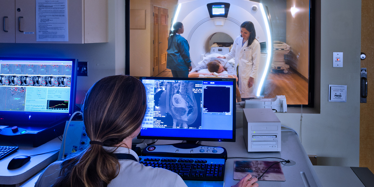
Focused Ultrasound Lab
Our research is focused on the development of new advances in MR imaging methodology, real-time data acquisition/reconstruction, and therapeutic ultrasound techniques. We have projects to develop MRI-guided focused ultrasound techniques for hyperthermia, high-intensity focused ultrasound (HIFU), and low-intensity focused ultrasound (LIFU). Research in those areas will provide promise for treating a wide range of oncological and neurological diseases.
Technical Developments
The lab develops advanced MRI-guided focused ultrasound technologies with an emphasis on accurate temperature monitoring, motion robustness, and real-time clinical integration.
- Motion-robust, multi-slice, real-time MR thermometry in abdominal organs
- Sonication strategies for volumetric ultrasound hyperthermia
- Temperature mapping robust to motion
- Real time integration (RTHawk) with InSightec system
- Temperature mapping in bone and fat
Basic and Clinical Research
MRI-guided focused ultrasound is being investigated as a powerful modality to treat a wide variety of conditions and applications, including movement disorders (such as essential tremor and Parkinson’s disease), brain tumors (such as glioblastoma and brain metastases), bone tumors (including osteoid osteoma), desmoid tumors, and psychiatric illness.
- Cardiac ablation with high-intensity ultrasound
- Prostate hyperthermia
- Pre-clinical studies:
- Bone regeneration after FUS
- Effect of sonication duration on ablation depth
- Facet joint ablation
- FUS-induced blood-brain barrier opening
Clinical Treatments and Trials
UCSF is consistently ranked among the nation’s top hospitals for the treatment of a wide variety of conditions. UCSF is a premier site for clinical trial enrollment and investigation because of strong collaborations between scientific and clinical enterprises. We are currently enrolling patients in clinical trials using focused ultrasound for the treatment of brain tumors, psychiatric illnesses, bone tumors, and others.
- Palliation of painful bone metastases through commercial and clinical trial
- Comparative Effectiveness of MRgFUS Versus CTgRFA for Osteoid Osteomas
- Treatment of desmoid tumors
- Focused Ultrasound to Promote Immune Responses for Undifferentiated Pleomorphic Sarcoma
- A Phase 1/2 Study of Sonodynamic Therapy Using SONALA-001 and Exablate 4000 Type 2 in Patients With DIPG
Research
This project proposed electronic beam steering, multi-focal patterns, and sector vortex beamforming approaches in conjunction with partial array activation.
This project proposed an effective and practical reconstruction pipeline to achieve motion-robust, multi-slice, real-time MR thermometry for monitoring thermal therapy in abdominal…
Feasibility of prostate hyperthermia with endorectal ultrasound applicators has been demonstrated in clinical trials
The goal of this study was to investigate near-field heating in patients treated with the InSightec fibroid system.
Proton resonant frequency shift (PRF) thermometry fails to detect temperature change in tissues with high lipid content, such as bone marrow.
Desmoid tumors are benign but locally aggressive soft tissue tumors that arise from fibroblast cells.
People
Focused Ultrasound Lab Researchers

Contact Us
Focused Ultrasound Lab
Matthew Bucknor, MD
Professor In Residence
[email protected]
Eugene Ozhinsky
Associate Professor
[email protected]