Program for Molecular Imaging and Targeted Therapy (Hope Lab)

The Program for Molecular Imaging and Targeted Therapy Lab involves a number of clinical trials focused on the translation of novel targeting imaging and therapy agents. We have a strong translation clinical research focus on novel radiotracers and targeted radionuclide treatments. We also work on clinical applications of simultinaneous PET/MRI. 

Research Directions

Our research lab has three main research focus:

PET/MRI

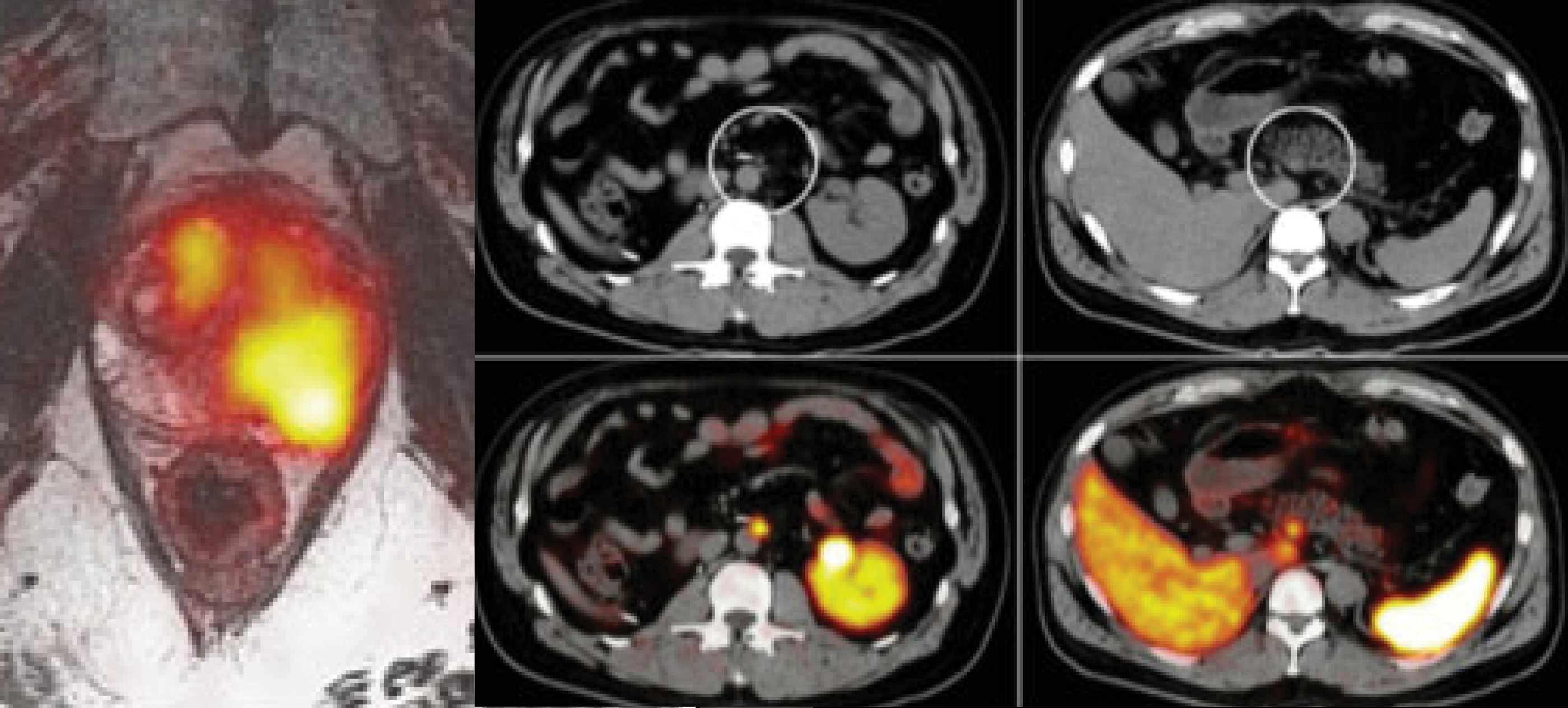
The simultaneous modality, PET/MRI, is a challenging environment due to the limitations of bringing PET and MRI together, but when appropriately leveraged, PET/MRI offers significant advantages over PET/CT.  Nonetheless, significant work remains to move the field forward, and we are working at UCSF to help address these:

  1. Imaging of pulmonary nodules: due to the short T2* of lung parenchyma, PET/MRI struggles in the detection of small pulmonary nodules compared to CT.  In oncology patients, pulmonary nodules are important and therefore many patients undergo an additional CT of the chest after imaging on a PET/MRI.  We are evaluating novel short echo pulse sequences (UTE and ZTE) to improve the detection of pulmonary nodules in the setting of PET/MRI.
  2. Improved approaches to attenuation correction: in the setting of PET/CT, attenuation correction is relatively straightforward as CT is a near direct measure of density.  In PET/MRI we cannot directly measure density and so other approaches have to be taken.  The major issue is in imaging bone, which has characteristics that make it difficult to imaging using PET/MRI.  Our initial work has been on using ZTE (zero echo time) MRI to allow for imaging of bone to improve attenuation correction maps for PET/MRI.
  3. Harmonization of quantitative accuracy: currently there are concerns about the quantitative accuracy of PET/MRI, primarily due to issues associated with attenuation correction.  Additionally there are no methods to qualify PET/MRI scanners for clinical trials.  Current phantoms used in PET/CT are not applicable for PET/MRI as they are not designed to be imaged using MRI.  We are working on creating a PET/MRI phantom and methodologies to harmonize quantification across MRI platforms.


Creation of a hybrid ZTE + Dixon spoiled gradient echo attenuation correction map to allow for improved quantification of osseous lesions.

Clinical Trials

  • Evaluation of Rectal Cancer Treatment Response Using PET/MRI (NCT02233595)

Publications

  • Yang J, Yiqiang J, Jenkins N, Behr SC, Hope TA, Larson PEZ, Vigneron D, Seo Y. Quantitative evaluation of atlas-based attenuation correction for Brain PET in an integrated Time-of-Flight PET/MR imaging system. Radiology. 2017.
  • Kluijfhout WP, Pasternak JD, Gosnell JE, Shen WT, Duh QY, Vriens MR, de Keizer B, Hope TA, Glastonbury CM, Pampaloni MH, Suh I. 18F Fluorocholine PET/MR Imaging in Patients with Primary Hyperparathyroidism and Inconclusive Conventional Imaging: A Prospective Pilot Study. Radiology. 2017 Jan 25:160768.
  • Leynes AP, Yang J, Shanbhag DD, Kaushik SS, Seo Y, Hope TA, Wiesinger F, Larson PE. Hybrid ZTE/Dixon MR-based Attenuation Correction for Quantitative Uptake Estimation of Pelvic Lesions in PET/MRI. Med Phys. 2017.
  • Hope TA, Verdin EJ, Bergsland EK, Ohliger MA, Corvera CU, Nakakura EK. Correcting for respiratory motion in liver PET/MRI: preliminary evaluation of the utility of bellows and navigated hepatobiliary phase imaging. EJNMMI Physics. 2015:2(1):21.
  • Burris NS, Johnson KM, Larson PE, Hope MD, Nagle SK, Behr SC, Hope TA. Detection of Small Pulmonary Nodules with Ultrashort Echo Time Sequences in Oncology Patients by Using a PET/MR System. Radiology. 2016 Jan;278(1):239-46.

Imaging and Treatment of Prostate Cancer

In the last few years there has been significant improvement in the imaging of prostate cancer using Prostate Specific Membrane Antigen (PSMA) PET.  We were the first institution in the United States to imaging patients using 68Ga-PSMA-11, otherwise known as the Heidelberg compound of HBED-CC PSMA.  We are involved in a multicenter trial to obtain data that will eventually lead to the FDA approval of the compound.  We are also progressing towards creating a trial using PSMA-617 as a targeted therapy treatment.


PSMA PET/MRI demonstrating uptake in a high grade primary tumor.  Diffusion weighted imaging (top right) and the associated ADC map (bottom right) demonstrated restricted diffusion, which correlates with the tumor seen on PSMA PET (bottom left).  Fused T2 weighted images (top right) demonstrates extracapsular extension.

Clinical Trials

  • Initial evaluation of PSMA PET in prostate cancer patients (NCT02611882)
  • Gallium-68 PSMA-11 PET in Intermediate to High-risk Preprostatectomy Patients (NCT0291911)
  • Gallium-68 PSMA-11 PET in Patients With Biochemical Recurrence (NCT02918357)

Publications

  • Hope TA, Truillet CC, Ehman EC, Afshar-Oromieh A, Aggarwal R, Ryan CJ, Carroll PR, Small EJ, Evans MJ. Imaging Response to Androgen Receptor Inhibition Using 68Ga-PSMA-11 PET: First Human Experience. J Nucl Med. 2016 Sep 22.
  • Afshar-Oromieh A, Hetzheim H, Kübler W, Kratochwil C, Giesel FL, Hope TA, Eder M, Eisenhut M, Kopka K, Haberkorn U. Radiation dosimetry of 68Ga-PSMA-11 (HBED-CC) and preliminary evaluation of optimal imaging timing. Eur J Nucl Med Mol Imaging. 2016 Aug;43(9):1611-20.

Imaging and Treatment of Neuroendocrine Tumors

Over the past five years I have worked on a number of projects related to neuroendocrine tumors.  We started by imaging patients with 68Ga-DOTA-TOC PET.  Currently we are running two trials using somatostatin receptor targeted therapies to treat patients.  We label compounds in house to offer unique treatments, and are studying different routes of administrations in order to increase treatment efficacy.


DOTA-TOC PET/CT in patient after resection of ileal primary, demonstrating focal uptake along the aorta (middle column) consistent with small residual disease.  Right column demonstrates physiologic uptake in the uncinate process.

Clinical Trials

  • Evaluation of Gallium-68 DOTA-TOC Imaging of Somatostatin Receptor Positive Malignancies (NCT02177773)
  • Expanded Access Protocol for Therapeutic Use of 177Lu-DOTA0-Tyr3-Octreotate in Patients With Inoperable, Somatostatin Receptor Positive, Midgut Carcinoid Tumors, Progressive Under Somatostatin Analogue Therapy (NCT02705313)
  • Yttrium-90 DOTA-TOC Intra-arterial (IA) Peptide Receptor Radionuclide Therapy (PRRT) for Neuroendocrine Tumor (pending)

Publications

  • Hope TA, Hernandez-Pampaloni H, Nakakura E, VanBrocklin H, Slater J, Jivan S, Aparici CM, Yee J, Bergsland E.  Simultaneous 68Ga-DOTA-TOC PET/MRI with gadoxetate disodium in patients with neuroendocrine tumor. Abdominal Imaging. 2015 Aug;40(6):1432-40.

Opportunities

Postdoctoral Position in Simultaneous PET/MRI

A postdoctoral appointment is available under the supervision of Dr. Peder Larson And Dr. Thomas Hope at the University of California, San Francisco (UCSF) Department of Radiology and Biomedical Imaging.  This position is focused on the development of tools to evaluate the quantitative accuracy of PET/MRI.  Work will involve PET reconstruction efforts, phantom development and work on novel MRI sequences to provide improved attenuation correction.  Position is fully funded for five years.

Learn more (docx)

People 

Professor
Vice Chair, Clinical Operations and Strategy
Associate Chair, Joint Ventures
Director of Molecular Therapy for MI&T
Professor In Residence
Professor
Director, Body RIG
Professor In Residence

Related Content