
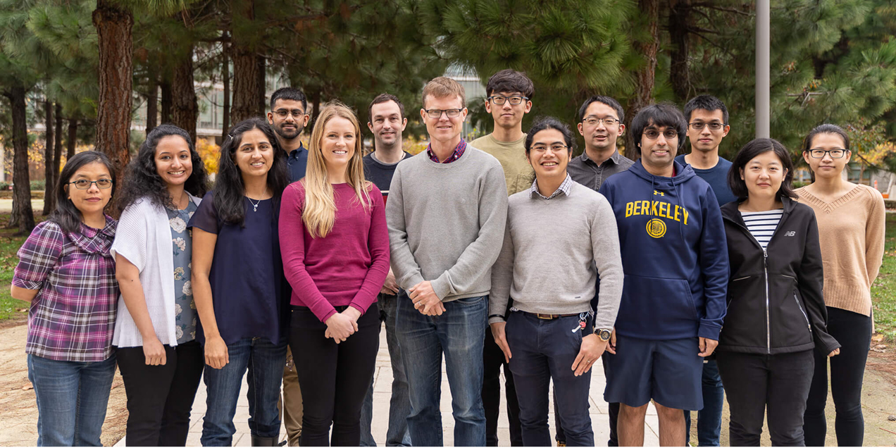
Larson Advanced Imaging Group
Our research group takes an engineering driven approach to developing advanced medical imaging methods, with a primary focus on MRI and additional work in CT and PET. The group is led by Professor Peder Larson and works closely with researchers and clinicians across UCSF. Our research emphasizes human and human ready imaging technologies and translational applications.
Research
We develop and apply methods in signal processing, optimization, signal modeling, statistical estimation, MRI scanner programming, and deep learning, with applications in human studies across oncology, urology, pulmonology, cardiology, and neurology.
Our group’s research projects span a range of advanced imaging technologies, including:
- AI methods for cancer imaging and prediction
- Hyperpolarized Carbon 13 MRI cardiac studies
- Hyperpolarized Carbon 13 MRI kidney studies
- Hyperpolarized Carbon 13 MRI methods
- Image reconstruction
- Lung MRI
- Metabolic MRI with hyperpolarized contrast agents
- Myelin imaging using ultra short echo time MRI
- PET MR and PET methods
- Radiation treatment planning
- Simultaneous PET MR imaging systems
Our team is based in Byers Hall at the UCSF Mission Bay campus as part of the Quantitative Biosciences Institute (QBI). Primary research facilities include 3T and 7T MRI systems, hyperpolarizers, and dedicated electronics and machine shops within the Surbeck Laboratory for Advanced Imaging, supported in part by the NIH funded Hyperpolarized MRI Technology Resource Center. We also conduct research on clinical imaging systems at UCSF hospitals and at the China Basin research facility, including 3T MRI scanners, a 0.55 T MRI scanner, and a time of flight PET MR system.
Additional resources, including a research blog, project descriptions, and software packages, are available on the Larson Advanced Imaging Group GitHub page
Opportunities
We are seeking strong PhD students and postdoctoral candidates. Please contact us if you are interested.
News
-
 Research
 -
 Education
 -
 Education
Educational Materials
We have developed and freely provide educational materials, primarily focused on MRI and hyperpolarized MRI, in the form of online textbooks, recorded lectures, and software, which are listed below.
- Introduction to Principles of MRI eBook by Peder Larson (and inspired by many, many others!)
- Hyperpolarized Carbon-13 Magnetic Resonance Imaging and Spectroscopy Edited by Peder Larson
- UCSF Biomedical Imaging 201: Principles of Magnetic Resonance Imaging (MRI)
- Lecture recordings: Principles of MRI - UCSF BI201 - YouTube
- UCSF Bioengineering 297: Hyperpolarized MR Seminar
- Several lectures as well as videos of Hyperpolarized MR experiments available at https://www.youtube.com/playlist?list=PLjBt5Iq93BT-cx76JwbQHwY1Y2nCJ30cS
- "MRI education resources" repository: https://github.com/LarsonLab/MRI-education-resources. (MATLAB based)
- Bloch simulations
- Image formation, RF pulse design
- Sample datasets
- eBook content
- Peder Larson - YouTube
- "Principles of (N)MR Imaging" Experimental NMR Conference, Educational Presentation, March 28, 2017.
- "Hyperpolarization - Description, Overview, & Methods" ISMRM Annual Meeting, Educational Presentation, April 26, 2017
- "Git Tutorial" UCSF, April 30, 2018
- "Lung MRI" ISMRM Annual Meeting, Educational Presentation, May 16, 2019
- "Software Development for Scientists" UCSF Practice of Science, February 18, 2020
- View: Slides
- "Mentoring Trainees in Research" UCSF Radiology Academic Research Series 2020
- View: Slides
- Educational Materials from Danish Research Centre for Magnetic Resonance. http://www.drcmr.dk/educations/education-material
- Introduction to MRI notes, targeted towards physicists and engineers
- Bloch equation and “compassMR” simulations for teaching (http://drcmr.dk/BlochSimulator/ http://www.drcmr.dk/CompassMR/)
- Videos explaining simulations
- MRI Course Lectures from Albert Einstein School of Medicine: https://www.youtube.com/playlist?list=PLPcImQzEnTpz-5TzxyyoYSbiAa9xdd89l
- Comprehensive series of lectures (56 in total) on MRI
- Starts from the basics, going into advanced pulse sequences and MR contrast
- Principles of Magnetic Resonance Imaging textbook by Dwight Nishimura. Available from lulu.com: Paperback or Hardcover
- Complete and coherent description of MRI, targeted towards engineers and physicists
- MRI From Picture to Proton textbook by McRobbie, Moore, Graves, and Prince, Cambridge University Press.
- Comprehensive description of MRI, targeted towards a less technical audience
- Many useful imaging examples and practical tips
- Questions and Answers in MRI http://www.mriquestions.com/index.html
- Answers to common questions about MRI
- Broad range, from the most basic to very advanced
- Bernstein, King, Zhou. Handbook of MRI Pulse Sequences. Academic Press. http://www.sciencedirect.com/science/book/9780120928613
- Detailed descriptions of specialized MRI topics
- Assumes introductory knowledge of MRI
- Essential for MRI scientists
- Schröder, Faber. In Vivo NMR Imaging. Spinger. https://link.springer.com/book/10.1007%2F978-1-61779-219-9
- Useful chapters on image formation, special contrast in MRI, and applications
- Very detailed descriptions
Lab Members
- Abhejit Rajagopal
Postdoctoral Scholar
Dr. Rajagopal received his PhD in 2019 from UC Santa Barbara under the mentorship of Shivkumar Chandrasekaran and Hua Lee. His thesis focused on the role of approximation theory in imaging and recognition algorithms. Since joining UCSF, Dr. Rajagopal has worked on machine learning techniques for enhanced PET/MRI reconstruction, prostate cancer grading, and techniques for quantifying generalization in deep learning. - Andrew Leynes, PhD
Bioengineering Graduate Student
Andrew Leynes is a graduate of the UCSF Masters of Biomedical Imaging Program and completed his PET/MR Master’s Thesis on “Tissue Segmentation and Classification for PET/MR MR-based Attenuation Correction using Zero-Echo Time (ZTE) MRI” in 2015. He is working on advanced methods for quantitative PET/MRI, high-field (7T) MRI, and RF coil design projects. - Anil Kemisetti
Associate Specialist
Anil Kemisetti received his master’s in Biomedical Imaging from UCSF in 2021. He also has a master’s in Health Informatics from the University of San Francisco and two decades of industry experience in software development. His work focused on Federated Learning. - Charlie Wang, MD, PhD
Radiology Resident
Dr. Charlie Wang was a Radiology Resident who completed his PhD at Case Western Reserve University on MR fingerprinting methods. He became an Assistant Professor in Radiology at UCSF. - Dharshan Chandramohan
Associate Specialist
Dharshan Chandramohan worked on quantitative cancer imaging methods using PET/MRI and to improve radiation therapy planning. He continued on to medical school in 2020. - Elizabeth Smith, PhD
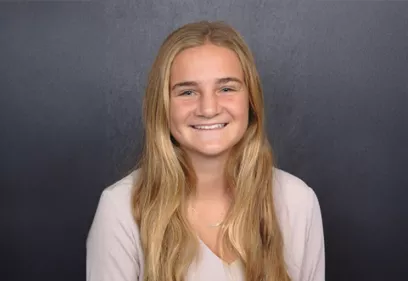
Data Science Fellow - Innovate for Health
Dr. Elizabeth Smith is a biophysicist and data scientist who is passionate about applying artificial intelligence and machine learning (AI / ML) to improve healthcare. Her current projects are focused on scaling the impact of PSMA PET in clinical decision making.
Dr. Smith spent the past five years at a geospatial analytics startup where she applied AI / ML to build software products from terabytes of satellite imagery. Her postdoctoral fellowship at UCSF and the Advanced Light Source focused on developing novel methods to image, reconstruct, co-align, and analyze features within three-dimensional tomographic reconstructions. She has a PhD in biophysics from the University of Wisconsin, Madison, and a bachelor's in physics from Pomona College. - Ernesto Diaz
Jr. Research Specialist
Mr. Diaz worked on data processing and analysis methods for hyperpolarized 13C MRI studies. - Fei Tan, PhD
Graduate Student
Dr. Fei Tan completed her PhD in Bioengineering from UC Berkeley - UCSF Bioengineering program. Her work focused on pulmonary ventilation analysis with ultrashort echo (UTE) proton MR. She is currently a Fellow at USFDA. - Jeremy Gordon, PhD
Postdoctoral Scholar & Senior Development Engineer
Dr. Jeremy Gordon came to UCSF from UW-Madison as a postdoctoral scholar in 2013 and he pioneered imaging methods and experimental protocols for human hyperpolarized carbon-13 metabolic MRI studies. He became an Assistant Professor at UCSF in 2020. - Jingwen Yao, PhD
Postdoctoral Scholar
Dr. Jingwen Yao completed her PhD in Bioengineering from the University of California Los Angeles under the mentorship of Dr. Benjamin Ellingson. Her dissertation involved developing and validating pH-sensitive chemical exchange saturation transfer (CEST) MRI in adult glioma patients. She worked on myelin imaging using ultrashort echo (UTE) relaxometry MRI with Dr. Peder Larson and multi-modal characterization of Huntington's disease (HD) at 7T with Dr. Janine Lupo. She is currently an Assistant Professor at UCLA. - Jessica Scholey, PhD
Bioengineering Graduate Student
Jess received her master's degree in Medical Physics and clinical residency training in Radiation Oncology from the University of Pennsylvania. She is currently a Board-Certified Medical Physicist and Bioengineering PhD student, where she focuses on MRI-applications in Radiation Oncology, specifically implementing sequence-based and deep learning-based approaches used for Radiotherapy dose calculation and incorporating these approaches into the clinical workflow. Jess is an Assistant Professor at UCSF Radiation Oncology. - Kirti Magudia, MD
Clinical Fellow
Dr. Magudia received her graduate training in the Weill Cornell / Rockefeller / Sloan-Kettering Tri-Institutional MD-PhD Program, where her PhD thesis focused on developing a 3D cell culture model of colon epithelial tumorigenesis. She completed her Radiology residency at Brigham and Women's Hospital, where she worked at the MGH & BWH Center for Clinical Data Science applying machine learning methods to define population-based normal values of body composition. She is currently an Assistant Professor at Duke University. - Manuska Vaidya, PhD
Postdoctoral Scholar
Dr. Vaidya received her PhD at the NYU School of Medicine under the mentorship of Ricardo Lattanzi, Graham Wiggins, and Dan Sodickson. She has extensive experience in RF coil design and evaluation of MRI evaluations, and is currently working on improved RF hardware and pulse sequences for brain tumor imaging with hyperpolarized carbon-13 MRI. - Naeim Bahrami
Biomedical Imaging Graduate Student
Naeim is a graduate of the the UCSF Master's of Biomedical Imaging program. He completed a Master’s thesis on “Modeling Hyperpolarized 13C Pyruvate and Urea Concentration Kinetics With Multibanded RF Excitation MRI In Prostate Cancer” in 2013, and went on to receive his PhD at Virginia Tech. - Ningjing (Nora) Zhang
Graduate Student
Ningjing (Nora) Zhang is a graduate student of the UCSF Master's of Biomedical Imaging program. She is currently working on her thesis “Improving Quantification of Prostate-Specific Membrane Antigen (PSMA) - Positron Emission Tomography (PET) Clinical Data via Deep Learning for Clinical Decision Making”. - Nick Dwork, PhD
Postdoctoral Scholar
Dr. Nicholas Dwork completed his PhD in Electrical Engineering from Stanford University with Prof. John Pauly working on acquisition and reconstruction of MRI and optical imaging data. He worked on novel methods for hyperpolarized C-13 MRI acquisition and reconstruction. Dr. Dwork is an Assistant Professor at the University of Colorado. - Peng Cao, PhD
Postdoctoral Scholar
Dr. Peng Cao was a postdoctoral scholar in the Larson Group at UCSF from 2014-2018, where he worked on a broad range of advanced imaging projects including advanced hyperpolarized carbon-13 MRS and MRI methods. He became an Assistant Professor at The University of Hong Kong in 2018. - Phil Yuan Tai Xie
Undergraduate Student
Phil Yuan Tai Xie is a UC Berkeley undergraduate student studying Electrical Engineering, Computer Science, and Bioengineering. Under the mentorship of Dr. Abhejit Rajagopal, Phil has been researching deep learning methods to improve multi-class cancer segmentation on MRI images. He studies the applications of latent space representations of brain and prostate cancer MRI images for improving the generalization performance of machine learning models. - Qing Dai
Biomedical Imaging Graduate Student
Qing is a graduate of the the UCSF Master's of Biomedical Imaging program. He completed a Master’s thesis on “Clear Cell Renal Cell Carcinoma: Deep Learning-Based Prediction of Tumor Grade from Contrast-Enhanced CT” in 2019, and worked in the group for one year before returning to UCLA to pursue his PhD. - Shuyu Tang
Bioengineering Graduate Student
Shuyu Tang was a graduate student in the UC Berkeley - UCSF Graduate Group in Bioengineering, and a graduate of the UCSF Master's of Biomedical Imaging program. He completed his thesis “Improved Acquisition Methods for Hyperpolarized Carbon-13 Magnetic Resonance Imaging” in 2019 and became an MRI research scientist in industry. - Sonam Machingal
Biomedical Imaging Graduate Student
Sonam is a graduate of the UCSF Master’s of Biomedical Imaging Program. She completed her Master’s thesis on “Sampling Strategies for Hyperpolarized Carbon-13 Dynamic Imaging” in 2014 under Dr. Larson. - Sule Sahin, PhD
Bioengineering Graduate Student
Dr. Sule Sahin completed her PhD in Bioengineering from the UC Berkeley-UCSF Graduate Group Bioengineering program. She worked on quantification and modelling of hyperpolarized carbon-13 imaging studies. - Tanguy Boucneau
Visiting Graduate Student
Tanguy Boucneau was a visiting graduate student in our group from the Ecole Normale Superior (ENS)-Cachan in France, working on ultrashort echo time (UTE) MRI methods for imaging myelin in the brain. He subsequently completed his PhD at the Université Paris-Saclay. - Wenwen Jiang
Bioengineering Graduate Student
Wenwen Jiang got her PhD in 2017 in the UC Berkeley - UCSF Graduate Group in Bioengineering, working jointly with Prof. Larson at UCSF and Prof. Michael Lustig at UC Berkeley on “Rapid and Robust Non-Cartesian Magnetic Resonance Imaging Methods”. Upon graduation, she became an imaging scientist in industry. - Xin Shen, PhD
Postdoctoral Scholar
Dr. Shen focused on sequence development, especially using rosette k-space trajectories for UTE MRI and MRSI, which has been demonstrated in myelin imaging, brain iron mapping, and other nuclei imaging including phosphorous and sodium. He is now working in Radiology at UCSD. - Xucheng Zhu
Bioengineering Graduate Student
Xucheng Zhu was a graduate student in the UC Berkeley - UCSF Graduate Group in Bioengineering. He worked on Motion Correction for lung imaging and dynamic hyperpolarized imaging strategies. He completed his thesis “Advanced 1H Lung MRI” in 2020 and became an MRI research scientist in industry. - Yan Ann Xing
Biomedical Imaging Graduate Student
Ann was in the inaugural class of the UCSF Master's of Biomedical Imaging program. She completed her Master's thesis on “Optimal Variable Flip Angle Schemes For Dynamic Acquisition Of Exchanging Hyperpolarized Substrates” in 2012 under Dr. Larson.
C13
- Christoffer Lausten - Aarhus University
- Jeremy Gordon
- Dan Vigneron - Vigneron Lab
- Zhen (Jane) Wang
- Duan Xu - Imaging Research for Neurodevelopment (Xu Lab)
- Renuka Sriram
- Yan Li
- Roselle Abraham
Lung
- Jae Ho Sohn
- Yang Yang
- Michael Lustig
- Kevin Johnson - University of Wisconsin
- Shreyas Vasanawala - Stanford University
- Xavier Maitre - Université Paris-Saclay
Neuro
- Roland Henry
- Ari Green
- Eduardo Caverzasi - University of Pavia
- Uzay Emir - University of North Carolina
PET/PET MRI
AI
- Sharmila Majumdar - Center for Intelligent Imaging
- Kirti Magudia - Duke University
- Zhen (Jane) Wang
- Tom Hope - Program for Molecular Imaging and Targeted Therapy (Hope Lab)
- Valentina Pedoia, PhD
- Corey Arnold - UCLA
- Antonio Westphalen - University of Washington
Radiation Oncology
- Allison Sabb, Summer Intern, Undergraduate Student
- Anna Bennett, Biomedical Imaging, Graduate Student
- Darren Hsu, Electrical Engineering and Computer Science, Undergraduate Student
- Dillon Yeh, Summer Intern, Undergraduate Student
- Eduarda Lopes, Summer Intern, High School Student
- Henry Salkever, Summer Intern, Undergraduate Student
- Jaelyn McKoy, Summer Intern, Undergraduate Student
- Jason Li, Summer Intern, Undergraduate Student, Boston University
- Jason Zhou, Electrical Engineering and Computer Science, Masters Student
- Josh Dean, Summer Intern, Undergraduate Student
- Jolie Wang, Summer Intern, High School Student
- Leila Abdelrahman, Summer Intern, Medical student
- Mario Quicana, Summer Intern, Undergraduate Student
- Rikhil Tanugula, High School Intern, Undergraduate Student
- Ritika Appanagari, Summer Intern, Undergraduate Student
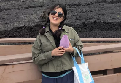
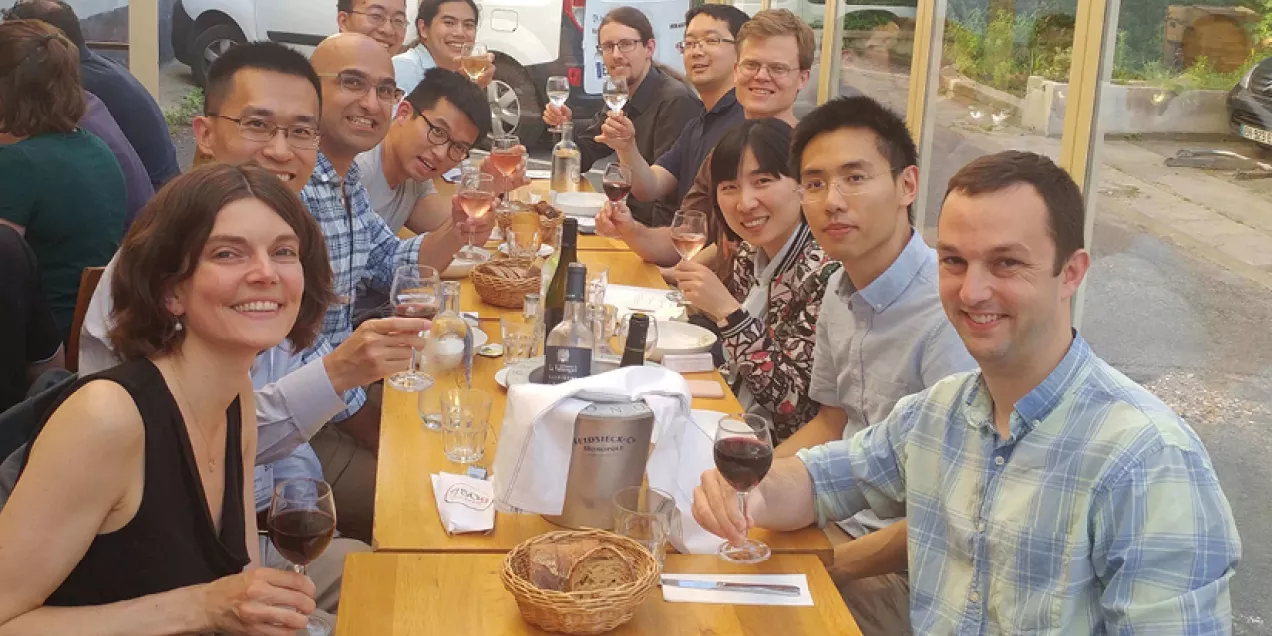
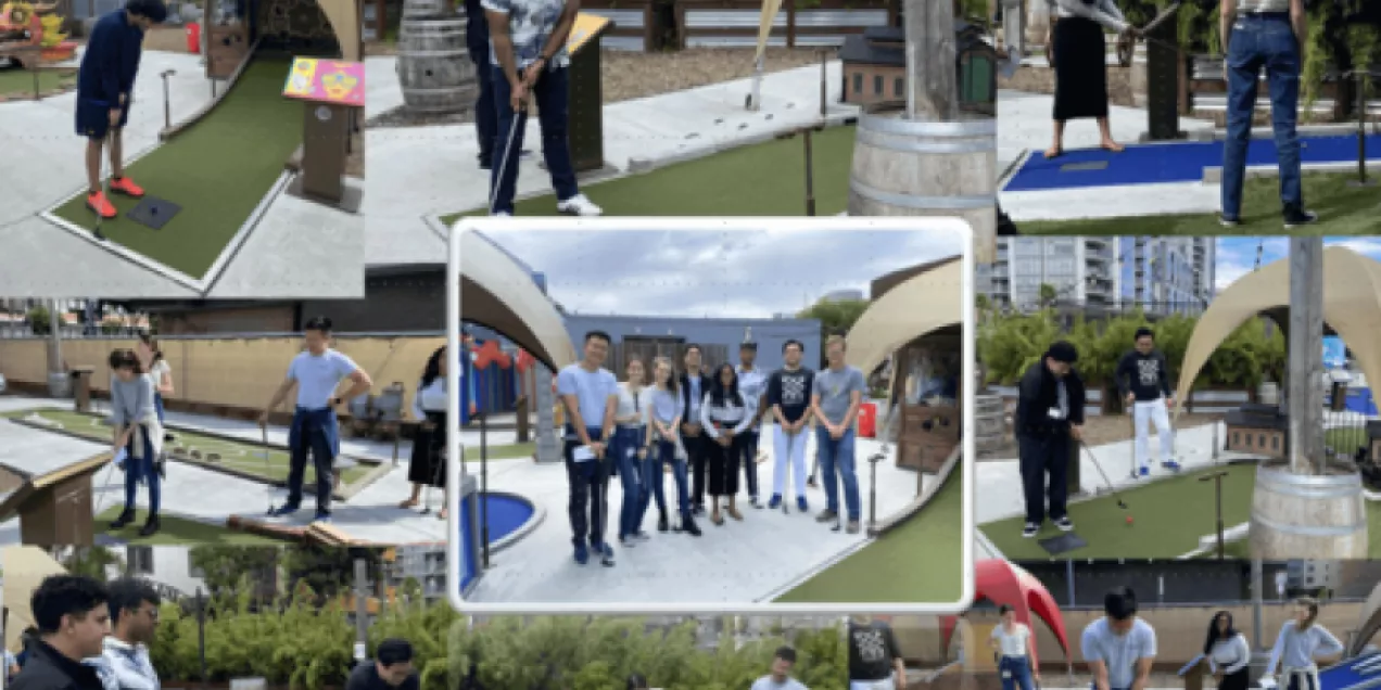
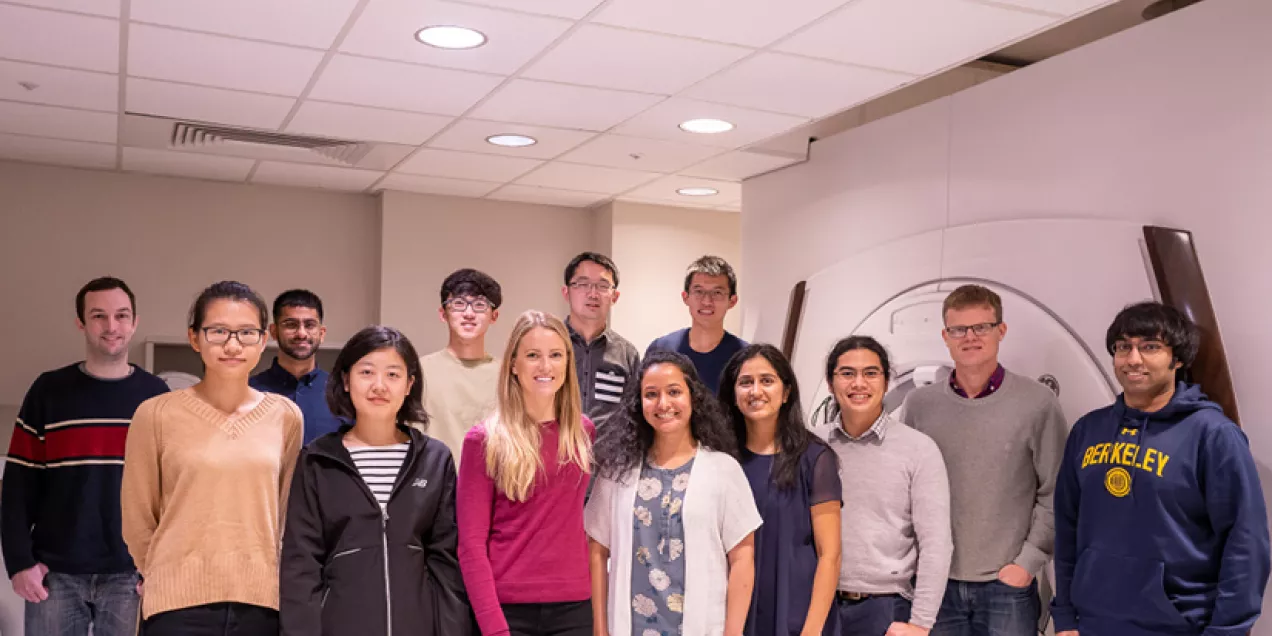
Lab Life & Team Moments
A collection of photos capturing our lab members over the years.
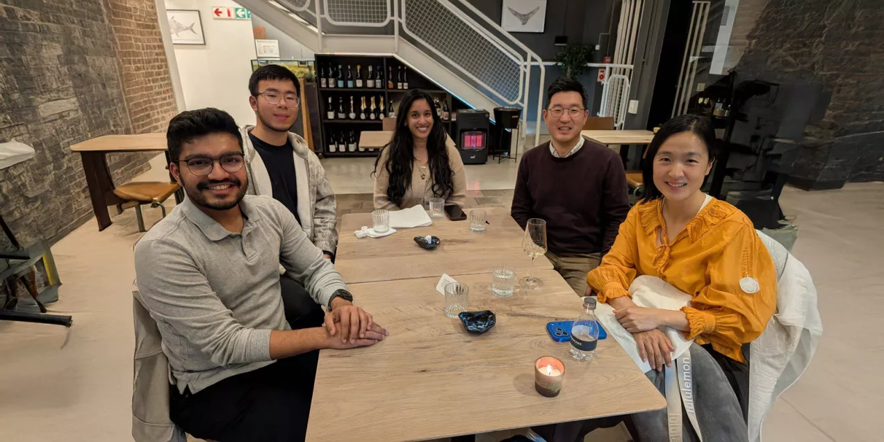
Lab Dinner at ISMRM 2026

Contact Us
Larson Advanced Imaging Group
Mission Bay Campus
1700 4th Street
Byers Hall, Suite 102
San Francisco, CA 94158
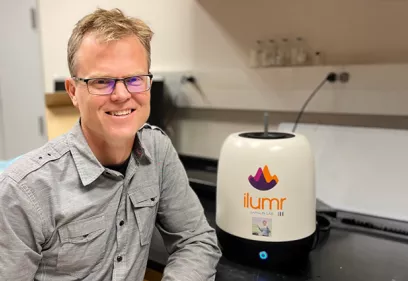
Peder Larson, PhD
Professor
Principal Investigator
Ph: (415) 514-4876
Fax: (415) 514-4451
[email protected]
Administrative Contact
Cresini Tabaranza-David
Ph: (415) 514-4450
[email protected]