Pediatric Radiology Clinical Research
Study of Advanced Magnetic Resonance Imaging for Pediatric Diseases (Dr. Jesse Courtier)
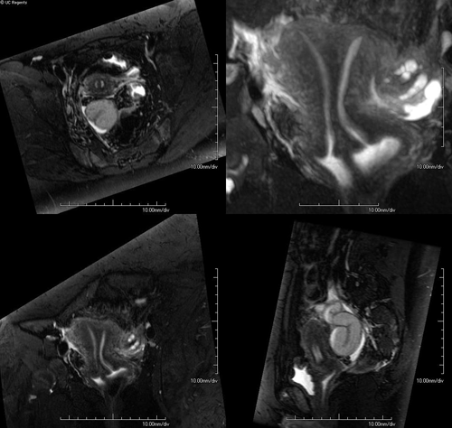
Dr. Courtier’s research focuses on the study of advanced non-invasive, radiation free imaging of children with magnetic resonance imaging (MRI). His research group studies diseases of the pediatric chest, abdomen and pelvis and how advanced imaging can better detect and monitor treatment of pediatric disease. Recent studies by his group include novel applications of MRI pulse sequences to improve the overall image quality and diagnostic capability of MRI. These include imaging techniques that are rapid and resistant to motion. A primary goal is to better visualize the small structures in children that often move and do this with less or no anesthesia. In addition, Dr. Courtier has a strong interest in resident and medical student education and research and has collaborated with students in a number of projects that have led to conference presentations and peer-reviewed publications.
Ongoing projects include:
- Study of novel diffusion weighted imaging (DWI) techniques to better identify inflammation and monitor treatment changes in children with inflammatory bowel disease (ulcerative colitis and Crohn disease).
- Testing and improving motion-resistant MRI with pulse sequences such as PROPELLER for use in infants and non-sedated children.
- Examining the strengths and limitations of high-resolution, 3D MRI sequences such as CUBE and SPACE for complex pediatric diseases in the chest, abdomen and pelvis including diseases of the liver, bile ducts, kidneys, and pelvic organs.
- Studying bowel motion (peristalsis) of bowel and the changes in motion that occur in disease using MRI pulse sequences such as real-time CINE FIESTA.
Dynamic MRI imaging of the bowel. Cine imaging is real-time imaging of the motion of the bowel. We are investigating this MRI technique for use in patients with inflammatory bowel disease and children with disorders of bowel motility.
Pediatric Molecular Imaging Laboratory (Dr. John MacKenzie)
Dr. MacKenzie’s primary research focus is molecular imaging. This new field of imaging shows promise in taking our understanding of disease and treatment monitoring to an entirely new level. Traditional clinical imaging examines how disease alters the anatomy of the body. Molecular imaging extends this anatomic imaging to an entirely new level. Molecular imaging helps us understand how diseases alter the function of life processes in the body. The field uses non-invasive imaging to detect how disease and therapeutic strategies influence molecular and cellular processes.
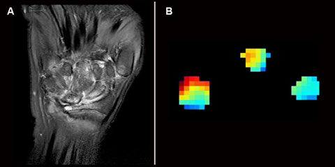
At the Pediatric Molecular Imaging Laboratory (PMIL) we study novel carbon-based molecular probes to better understand and treat childhood disease. Currently we would like to better understand inflammatory arthritis (rheumatoid and juvenile idiopathic arthritis) using high-field MRI. Dr. MacKenzie and his team test new molecular imaging strategies and use several preclinical modes of inflammation. The quantitative, objective, and noninvasive biomarkers underdevelopment in the PMIL may help better detect and monitor treatment of these diseases.
Pediatric Imaging Education and Advanced Ultrasound (Dr. Andrew Phelps)
Dr. Phelps research focuses on the development of new methods for clinical education. Dr. Phelps is an active member of Association of University Radiologists and has authored six peer-reviewed publications focused on improving the training of radiology students: medical students, residents, and fellows.
Dr. Phelps also is an accomplished medical illustrator. The medical community recognizes his illustrations as valuable teaching tools that help convey important concepts. His illustrations are widely present in formal lecture materials, medical journals and medical textbooks.
Dr. Phelps’ experience with pediatric ultrasound has also been put to good use in teaching anatomy to UCSF medical students. Dr. Andrew Phelps complements his special interest in medical illustration with anatomy education and uses ultrasound when he teaches anatomy to medical students. The ability for medical students to use ultrasound to peer into their own anatomy has been a successful complement to their traditional cadaveric dissection.


