Clinical & Translational Musculoskeletal Imaging
Research Projects
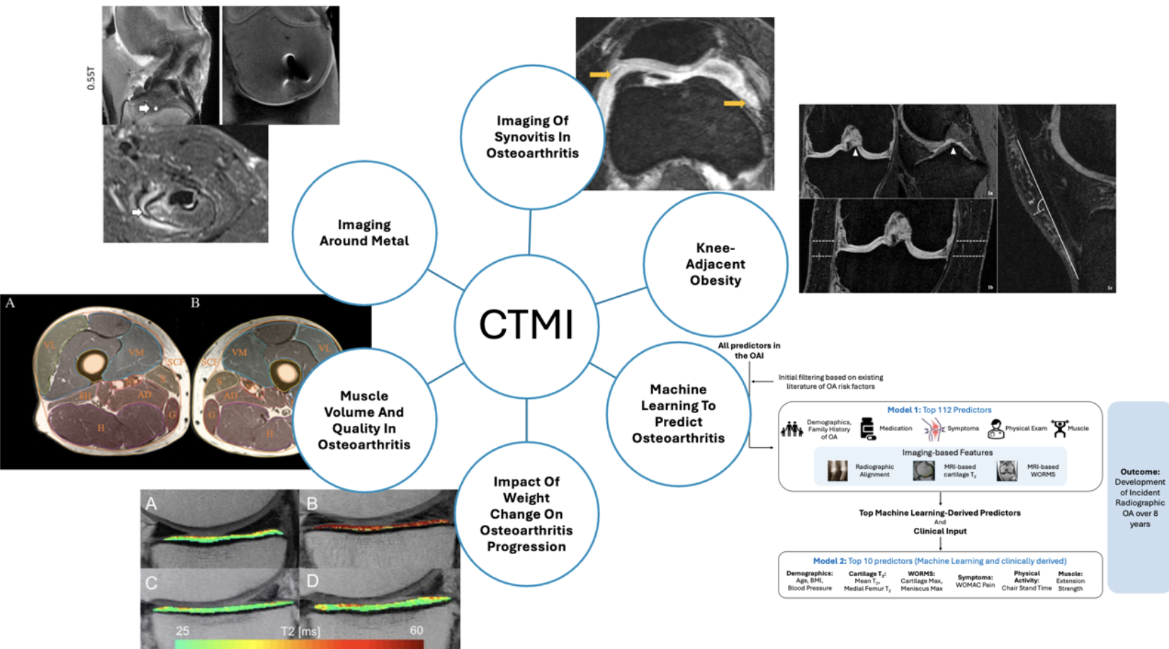
Imaging of Synovitis in Osteoarthritis
Our team found that greater progression of structural degenerative disease of the knee was observed in individuals with sustained synovitis (joint inflammation) compared to those without sustained synovitis, suggesting that sustained synovitis is associated with progressive osteoarthritis.
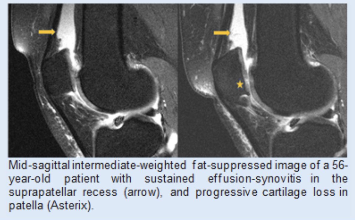
We also found that effusion-synovitis progression was slowed by weight loss and decrease in local subcutaneous fat. Hoffa-synovitis characterized by fluid in the infrapatellar fat pad increased at the same time, suggesting a decreasing fat pad rather than active synovitis. Decrease in local subcutaneous fat partially mediated the systemic effect of weight loss on synovitis.
Read more about these projects:
Impact of Sustained Synovitis on Knee Joint Structural Degeneration: 4-Year MRI Data from the Osteoarthritis Initiative. Ramezanpour S, Kanthawang T, Lynch J, McCulloch CE, Nevitt MC, Link TM, Joseph GB. J Magn Reson Imaging. 2023 Jan;57(1):153-164. doi: 10.1002/jmri.28223. Epub 2022 May 13.
Effect of weight loss on knee joint synovitis over 48 months and mediation by subcutaneous fat around the knee: data from the Osteoarthritis Initiative. Löffler MT, Ngarmsrikam C, Giesler P, Joseph GB, Akkaya Z, Lynch JA, Lane NE, Nevitt M, McCulloch CE, Link TM. BMC Musculoskelet Disord. 2024 Apr 17;25(1):300. doi: 10.1186/s12891-024-07397-y.
Impact of Weight Change on Osteoarthritis Progression
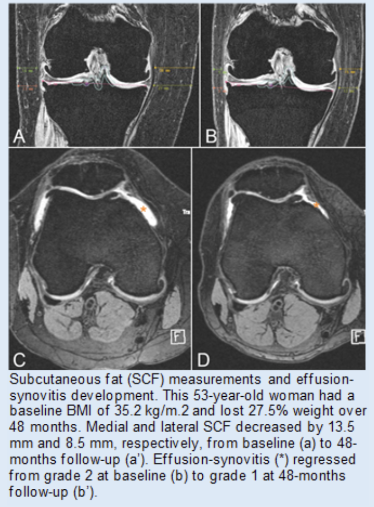
We investigated the effects of weight loss and weight gain on hip and knee radiographic changes, pain, and joint replacement over 4 years in the Osteoarthritis Initiative Cohort. Our results of this large, longitudinal study (n = 2,752 with 4-year follow-up) suggested that weight loss may protect against, and weight gain may exacerbate, radiographic and symptomatic knee OA, while weight change (at a 5% threshold) does not have significant effects on hip OA.
Read more about these projects:
Effects of Weight Change on Knee and Hip Radiographic Measurements and Pain Over Four Years: Data From the Osteoarthritis Initiative. Joseph GB, McCulloch CE, Nevitt MC, Lynch J, Lane NE, Link TM. Arthritis Care Res (Hoboken). 2023 Apr;75(4):860-868. doi: 10.1002/acr.24875. Epub 2022 Nov 18.
Associations between weight change, knee subcutaneous fat and cartilage thickness in overweight and obese individuals: 4-Year data from the osteoarthritis initiative. Joseph GB, Takakusagi M, Arcilla G, Lynch JA, Pedoia V, Majumdar S, Lane NE, Nevitt MC, McCulloch CE, Link TM. Osteoarthritis Cartilage. 2023 Nov;31(11):1515-1523. doi: 10.1016/j.joca.2023.07.011. Epub 2023 Aug
The effect of interactions between BMI and sustained depressive symptoms on knee osteoarthritis over 4 years: data from the osteoarthritis initiative. Joseph GB, McCulloch CE, Nevitt MC, Lynch J, Lane NE, Pedoia V, Majumdar S, Link TM. BMC Musculoskelet Disord. 2023 Jan 12;24(1):27. doi: 10.1186/s12891-023-06132-3.
Role of Ultra-Processed Foods in Knee Joint Health & Muscle Quality
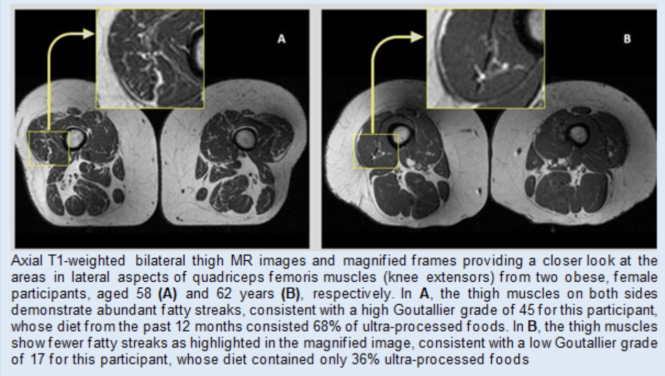
In this project, we found that the more ultra-processed foods people consumed, the more intramuscular fat they had in their thigh muscles, regardless of caloric intake or physical activity. We hypothesize that consuming ultra-processed foods, such as cereals, frozen meals, soft drinks, and packaged snacks, may also raise knee osteoarthritis risk. This was the first imaging study looking into the relationship between skeletal muscle quality and quality of diet.
RSNA press release: Eating High-Processed Foods Impacts Muscle Quality
Media Coverage:
Machine Learning to Predict Osteoarthritis
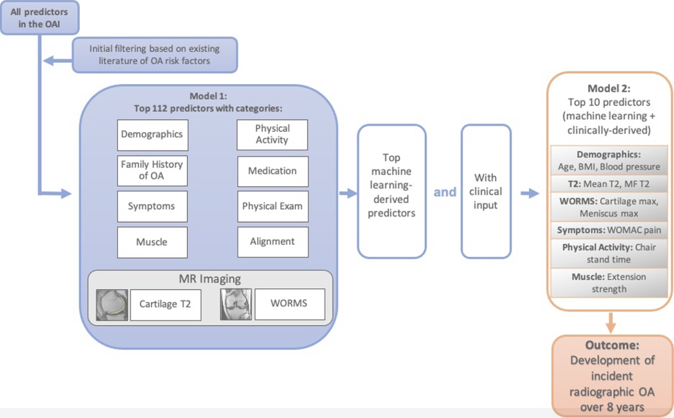
We developed a machine learning-based prediction model for incident radiographic osteoarthritis (OA) of the knee over eight years using MRI-based cartilage biochemical composition and knee joint structure, demographics, and clinical predictors, including muscle strength and symptoms. We found that a 10-predictor model including MRI parameters coupled with demographics, symptoms, muscle, and physical activity scores provided good prediction of incident radiographic OA over eight years.
Read more about this project:
Machine learning to predict incident radiographic knee osteoarthritis over 8 Years using combined MR imaging features, demographics, and clinical factors: data from the Osteoarthritis Initiative. Joseph GB, McCulloch CE, Nevitt MC, Link TM, Sohn JH. Osteoarthritis Cartilage. 2022 Feb;30(2):270-279. doi: 10.1016/j.joca.2021.11.007. Epub 2021 Nov 18.
Knee Adjacent Obesity
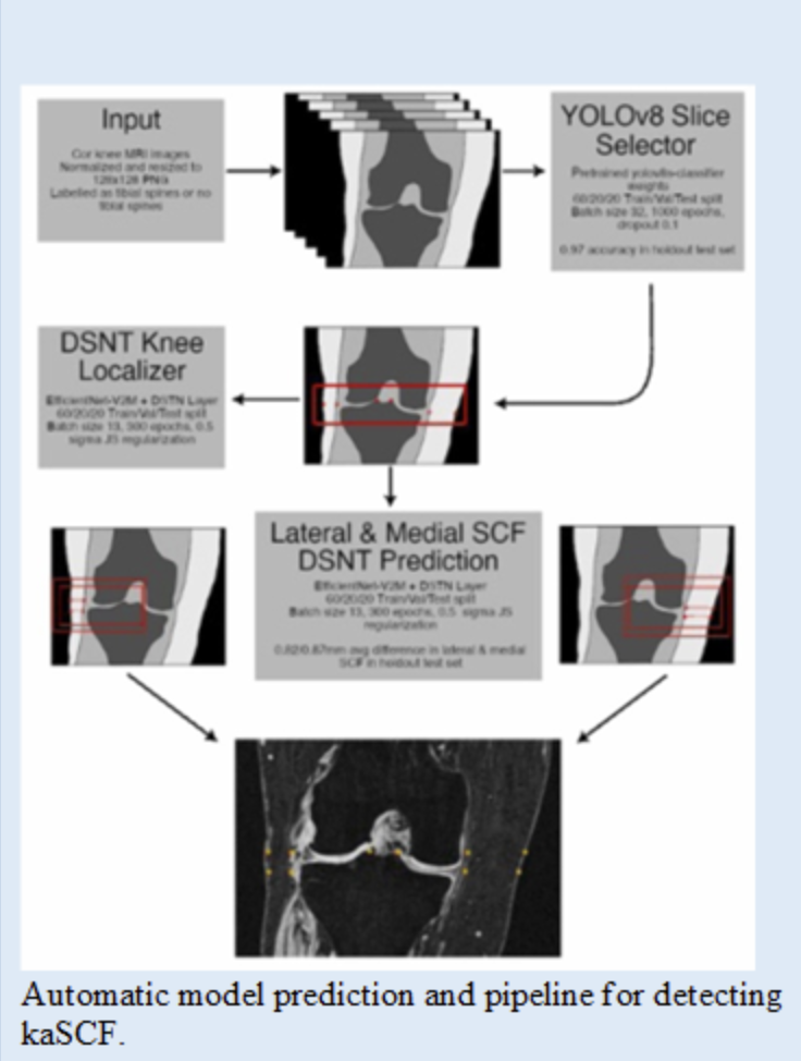
Knee-adjacent subcutaneous fat (kaSCF) has emerged as a potential biomarker and risk factor for osteoarthritis (OA) progression. We developed an artificial intelligence-based tool for the automatic segmentation of kaSCF thickness and evaluated the cross-sectional associations between kaSCF, cartilage thickness, magnetic resonance imaging-based cartilage T2 relaxation time, knee pain, and muscle strength independent of body mass index (BMI). We found that greater kaSCF was associated with thinner cartilage in men, higher T2 in women, reduced knee strength, and greater knee pain, independent of BMI. These findings suggest a potential role of kaSCF as a predictor for knee osteoarthritis-related structural, functional, and clinical outcomes independent of the effects of BMI.
Read more about this project:
Quantifying knee-adjacent subcutaneous fat in the entire OAI baseline dataset - Associations with cartilage MRI T2, thickness and pain, independent of BMI. Joseph GB, Liu F, Ziegeler K, Akkaya Z, Lynch JA, Pedoia V, Majumdar S, Lane NE, Nevitt MC, McCulloch CE, Link TM. Osteoarthritis Cartilage. 2025 Apr;33(4):482-490. doi: 10.1016/j.joca.2025.01.001. Epub 2025 Jan 27.
Muscle Volume & Quality in Osteoarthritis
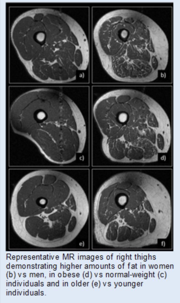
The degree of thigh intramuscular fat in individuals without OA is fundamental for distinguishing natural variations in intramuscular fat from pathological changes. In this project, we investigated the degree of thigh intramuscular fat in individuals without radiographic OA or frequent pain and assessed the associations of age, sex, and BMI with the degree of intramuscular fat. Goutallier Grades (GGs) of the quadriceps and hamstring muscles were assessed based on 3 T MR images on a scale from 0 (normal muscle) to 4 (more fat than muscle). The associations between demographic variables and GG outcomes were evaluated using mixed effects models. While individuals without radiographic OA or frequent pain generally had low thigh intramuscular fat, higher BMI and age were associated with greater intramuscular fat, and GGs were greater in women than men. The relationship between BMI and intramuscular fat was sex-dependent. Thus, demographic variables must be considered when evaluating intramuscular fat.
Read more about this project:
Thigh muscle and fat volumes are associated with knee cartilage abnormalities and bone marrow edema-like lesions: data from the osteoarthritis initiative. Manatrakul R, Pirmoazen AM, Bharadwaj UU, Akkaya Z, Giesler PJ, Lynch JA, Nevitt MC, McCulloch CE, Joseph GB, Link TM. Skeletal Radiol. 2024 Jul;53(7):1279-1286. doi: 10.1007/s00256-024-04565-y. Epub 2024 Jan 11. PMID: 38206355
MRI-based analysis of thigh intramuscular fat and its associations with age, sex, and BMI using data from the osteoarthritis initiative data. Joseph GB, Akkaya Z, Sims WM, McCulloch CE, Nevitt MC, Lynch JA, Lane NE, Link TM. Sci Rep. 2025 Feb 20;15(1):6188. doi: 10.1038/s41598-024-75005-z.
Imaging Around Metal
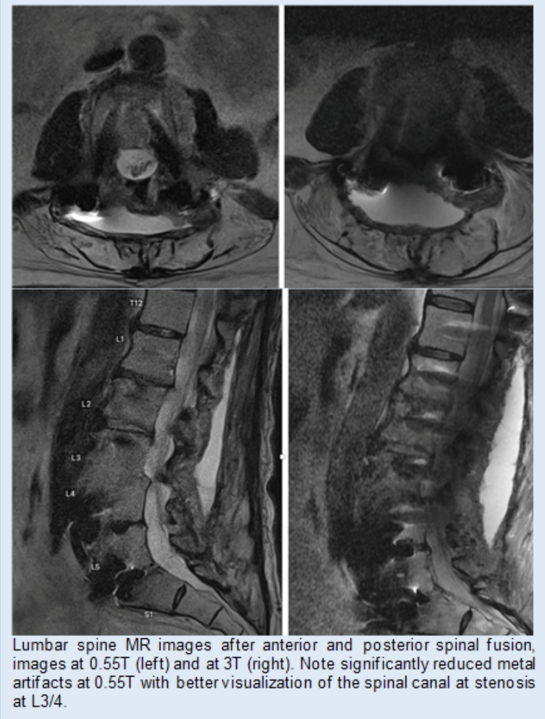
Using a novel 0.55T system we studied patients with metal implants at the spine (spinal fusion with metal hardware) and hip (hip arthroplasties) and found that imaging at 0.55T had superior image quality and showed abnormalities better than imaging at 1.5 or 3T. We also performed a biophantom study using metallic implants (steel and titanium screws) in pig knees which demonstrated substantial reduction of artifact size resulting in superior depiction of anatomical structures at 0.55 T MRI.
Read more about this project:
Improved metal suppression using new generation low-field MRI: a biophantom feasibility study. Luitjens J, Ziegeler K, Yoon D, Gassert F, Bhattacharjee R, Manatrakul R, Ngarmsrikam C, Becker A, Yang Y, Joseph GB, Su P, Itriago-Leon P, Majumdar S, Link TM. Skeletal Radiol. 2025 May;54(5):1093-1099. doi: 10.1007/s00256-024-04809-x. Epub 2024 Oct 4.
Publications
See a complete list of the group’s publications at Dr. Link’s UCSF profile.