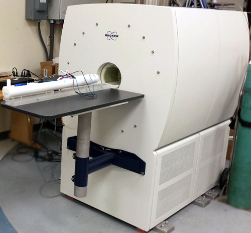
3T MRI at NMR Lab

The Bruker 3T scanner is specially designed for pre-clinical MRI studies of rats and mice. A special dual tuned 13C-1H RF coil is available for hyperpolarized DNP studies using 13C labelled biomarkers. The scanner is equipped with a cryogen free magnet and high performance (90G/cm) gradients.
Getting Started

We provide you with full support along all aspects of your study, from experiment and protocol design, to animal care and housing, to image analysis and data management.  Beginning the process is easy; you can start by contacting us with questions and preliminary ideas for your experiment, and then take a look at our online  study application.

Equipment


The Bruker 3T scanner is specially designed for pre-clinical MRI studies of rats and mice. A special dual tuned 13C-1H RF coil is available for hyperpolarized DNP studies using 13C labelled biomarkers. The scanner is equipped with a cryogen free magnet and high performance (90G/cm) gradients.

Imaging Capabilities and Sequences

• Anatomic Imaging (T1/T2/ Proton-Density weighted imaging)
• Different T1, T1rho, T2 and T2* mapping methods
• Diffusion Weighted Imaging (DWI), Diffusion Tensor Imaging (DTI) and Diffusional Kurtosis Imaging (DKI)
• Blood Flow and Blood Oxygen Measurements (BOLD EPI methods)
• Angiography using Contrast Agents or Time Of Flight (TOF)
• Fat/Water imaging based on spin- or gradient-echo imaging (Dixon techniques)
• 2D and 3D Chemical Shift Imaging (CSI)
• Metabolite Editing Methods (Lactate detection)
• Single Voxel Spectroscopy (LASER /PRESS)
• SWIFT Imaging
• Magnetization Transfer and CEST imaging techniques
• Dynamic Contrast Enhancement with Gadolinium
• Manganese Enhanced Imaging
• B0/B1 Mapping techniques
• Multi-Modal Imaging (CT, Optical, and PET/CT)
• Sequence/Protocol Design Services Available

Data Analysis and Management

The 7T Small Animal MRI is integrated with the Quantitative Image Processing Center (QUIPC), to provide start-to-finish image analysis and data management.  

  • Pre- and Post-Processing
  • Segmentation
  • Co-Registration
  • Quantitative Parametric Maps (T1, T2, DCE, BMD, etc.)
  • Diffusion Tensor Analysis
  • Custom Analysis Software Development

Rates: 3T MRI Intramural/Extramural

Core


Rate Description Unit Internal Rate External Rate Rate Change Effective Date Internal Rate Change Pending External Rate Change Pending
Pre-clinical Imaging Core Services Scan Rate hour $118.00 148.68      
Technician Support hour $67.00 $84.42      
HyperSense Polariser sample $72.00 $90.72      
Pass through expense   at cost at cost + 26%      

EXAMPLE DATA

Parameter Mapping

7T
Diffusion and T2 mapping of brain tumor models (mouse)
Left-hand image: Apparent Diffusion Coefficient; Right-hand image: T2

Functional

TAGCINE of Mouse Heart 


Manganese Enhanced MRI of Mouse Olfactory Activation


Metabolic Editing Methods (Lactate Detection)


Metabolic imaging of brain tumor models
Left-hand image: T2 weighted reference; Right-hand image: Lactate overlay map

Whole Mouse Body Lipid Imaging (in vivo)


Lipid distribution (mouse)

Perfusion Imaging using FAIR-ASL


Perfusion imaging of the rat brain

Stroke Imaging


Stroke imaging (rat pubs) using diffusion weighted imaging

Diffusion Tensor Imaging (DTI)


DTI imaging of the rat spinal cord using reduced FOV EPI

Anatomical Imaging


Anatomical imaging of the spinal cord (mouse)

Localized Spectroscopy


Localized spectroscopy of the mouse brain using the LASER technique

Cartilage Imaging


Cartilage imaging of the rat knee

fMRI of the Rat Brain Using rFOV EPI Imaging


Left-hand image: T2-weighted Turbo-Spin-Echo image; Right-hand image: rFOV Single-Shot-EPI image

Fat/Water Imaging (Mouse Shoulder, ex vivo)


Left-hand image: Water distribution; Right-hand image: Lipid distribution

Magnetic Resonance Angiography (MRA)


Contrast enhanced MRA of the mouse brain
Left-hand image: Control; Right-hand image: AVM-Model

Diagnostic/Anatomical Imaging


Imaging of lung tumors (mouse)

High-Resolution Diffusion Tensor Imaging


DTI imaging of the rat heart (ex vivo)
Left-hand image: Eigenvector; Right-hand image: Mean diffusivity