Nuclear Medicine - Clinical Nuclear Imaging Research (C-NIR)

 The C-NIR group is dedicated to performing Molecular Imaging Research in humans with state of the art PET/CT and SPECT/CT capabilities. Our imaging scanners are run by highly trained and qualified Nuclear Medicine and CT certified technologists. We also have imaging science experts to provide additional help on diagnosis, image reconstruction, co-registration, tracer kinetic modeling, and quantifications.
We provide asistance with all aspects of your study including experiment and protocol design, patient imaging and data analysis. Please contact us with your questions or ideas.
Getting Started
 A "research study" PET/CT or SPECT/CT consist of any PET/CT or SPECT/CT performed in humans that differs in some fashion from the standard clinical protocol. The deviation from the standard clinical protocol could be (but not limited to): the radiopharmaceutical, the acquisition protocol, the reconstruction or the biodistribution time.
Five main things are needed to get a research study in human subjects started with us (not to be obtained necessarily in this order):
- A research imaging protocol. We provide consultation and can help you develop your research idea into an imaging protocol.
- A RUA (Radioisotopic User Authorization) approval from the UCSF RSC (Radiation Safety Committee)
- Approval from the UCSF IRB committee
- Fill out and submit the Full Study Application form. This will make sure we can accommodate your study from an operational point of view, to make your study happen.
- Have the study reviewed and approved by the CNIR committee, which meets at the beginning of every month. Once approved, our CRC will contact you and will be ready to help with study related matters such as patient scheduling.
Capabilities

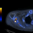
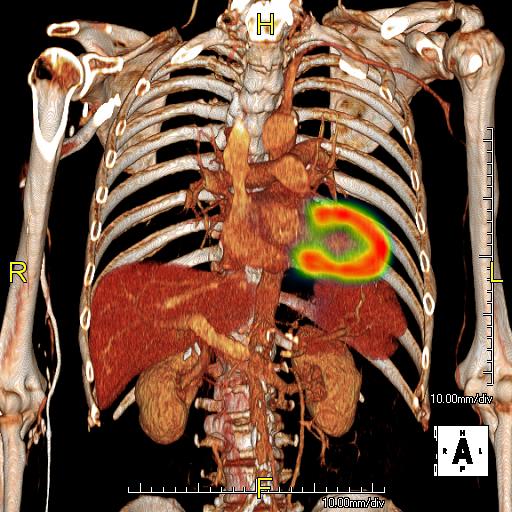
- PET/CT Imaging
- SPECT/CT Imaging
- Radio Pharmaceutical
- Radionuclide Therapy
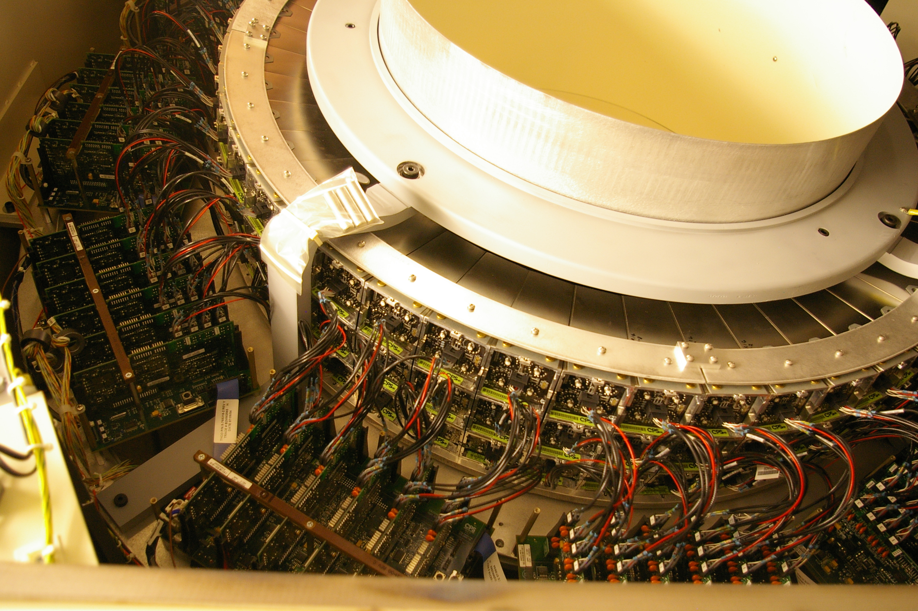
Equipment
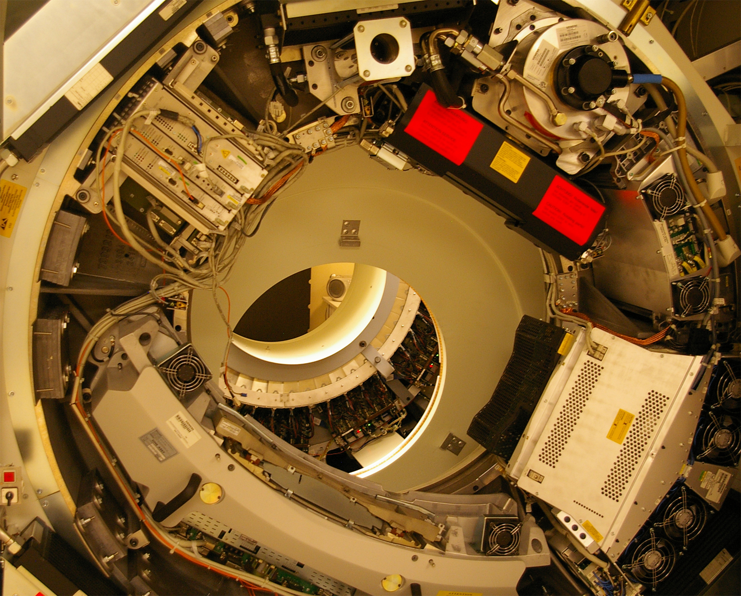
UCSF China Basin Imaging Center
- GE Discovery VCT PET/CT
- Siemens Biograph 16 PET/CT
- GE Infinia Hawkeye 4 SPECT/CT
- GE Healthcare Signa 3.0T Time-Of-Flight PET/MRI
UCSF Moffitt/Long Hospital
- GE Discovery NM/CT 670
- Siemens Exact HR+ PET
Radiopharmaceuticals Available at UCSF
 PET Radiopharmaceutical Status
Fludeoxyglucose F 18 Injection | Active Ammonia N 13 Injection | Active Sodium Fluoride F 18 Injection| Active O-15 Water | Active F-18 FMISO | Active F-18 FLT NEPTIS method | Active F-18 FLT NCI method | ActiveC-11 DHE | ActiveI-124 MIBG | Active F-18 Florbetapir (AmyvidTM) | Active C-11 Methoinine | Inactive other commerically available FDA approved radiopharmaceutical agents - ex: RB-82, DaTSCAN (123I-Ioflupane)
Active=available for clinical use; Inactive=requires qualification runs for clinical use
Standard PET/CT Protocols
Standard Body PET/CT:
- Uptake Time – 60 minutes
- Patient Positioning – Head first, supine with arms above the head (for optimal thoracic imaging).
- Scan Length – Top of skull to the mid-thigh
- Scan Duration – 20 - 30 minutes (generally 8 - 10 bed positions)
- Scan Direction – From head toward feet
Whole Body PET/CT:
- Uptake Time – 60 minutes
- Patient Positioning – Head first, supine with arms crossed low over the abdomen (allows for scanning of the entire body including arms)
- Scan Length – Top of the head to the tip of the toes (usually done as a single scan, but may be split into 2 scans for tall people)
- Scan Duration – 35 - 50 minutes
- 1.5 minutes per bed position for the body and 40 seconds per bed position for the legs for patients with BMI < 25
- 2 minutes per bed position for the body and 80 seconds per bed position for the legs for patients with BMI between 25 and 30
- 3 minutes per bed position for the body and 120 seconds per bed position for the legs for patients with BMI ≥ 30
- Scan Direction – From head toward feet
Standard Brain PET/CT:
- Uptake Time – 30 minutes
- Patient Positioning – Head first, supine with arms crossed over the chest/abdomen or at sides (for patient comfort)
- Scan Length – Single bed position that includes the entire brain
- Scan Duration – 10 minute single time frame
- Scan Direction – Single bed position