Preclinical MR Imaging and Spectroscopy Lab
Discovery and Clinical Translation of Imaging Biomarkers
The Preclinical MR Imaging and Spectroscopy Laboratory is located in Genentech hall, the building adjacent to the Quantitative Bioscience Institute (QBI), on the UCSF Mission Bay Campus. This state-of-the-art Core facility offers MRI and MRS capabilities which are uniquely integrated with DNP polarizers for conducting hyperpolarized 13C MR experiments. All magnets are dedicated to running biomedical samples and they have complementary features, including high-resolution magic angle spinning (HR-MAS) spectroscopy, micro-imaging enabling NMR studies of biopsy, surgical tissues, cell and tissue cultures and murine models of cancer and other diseases, and in vivo hyperpolarized 13C studies in rodents as well as in perfused cell and tissue bioreactors. The details of each system can be found below.
If you are interested in using the Preclinical MR Imaging and Spectroscopy Lab Core facility for your studies, please contact Renuka Sriram, PhD (Technical Director) for more information.

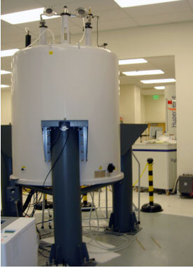
 The 14.1 Tesla Agilent wide-bore micro-imaging NMR spectrometer is equipped with 100mT/m gradients. It has excellent cardiac and respiratory gating performance and has demonstrated the ability to obtain high-resolution 1H and hyperpolarized 13C MRSI data in mice, in multiple organs (brain, prostate, liver, etc...). In close collaboration with Agilent Instruments Inc. (Palo Alto, CA), the imaging spectrometer was custom designed to accommodate the technical requirements of performing 1- and 2-D multinuclear spectroscopic studies of tissues, cultured cells and tissue bioreactor studies, and micro-imaging and hyperpolarized spectroscopic imaging studies. Specifically, the imaging spectrometer enables: 1) the acquisition of 1H 2D and multinuclear HR-MAS spectra from small samples (6-10 mg), such as image-guided biopsies, in a short enough time to avoid pathologic, metabolic and RNA degradation; 2) the ability to perform ex vivo and in vivo 3D anatomic, perfusion, diffusion MRI and spectroscopic imaging studies of cultured tissues and cells, and mouse models; and 3) the ability to utilize novel hyperpolarized 13C labeled substrates and 13C spectroscopic imaging techniques to identify exciting new biomarkers. Furthermore, an isoflurane-based inhalant anesthesia system is located next to the system for preclinical studies. Thermal support for animals during preparation and imaging is provided by a combination of heat lamps and heated thermal water pads (with deuterated or Mn-doped water during imaging), and temperature-regulated hot air fans according to individual project needs. A kit (SA Instruments, Inc.) allowing rectal thermometry, ECG probes/amplifier with TTL trigger for external gating, and respiratory pick-up is also available for continuous monitoring of the animal's physiological parameters.
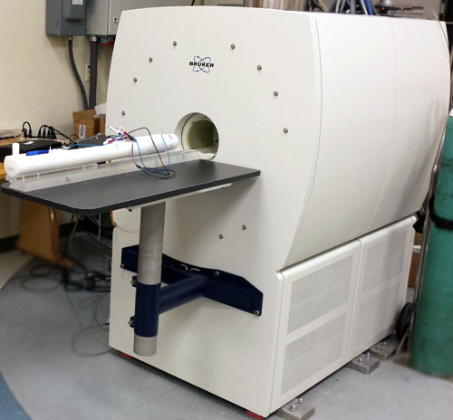
 Especially designed for study of mice and rats with a magnet bore of 180mm, the BioSpec 3T comprises the latest Bruker MRI technology, Paravision software application packages and multimodal options at the translational field of 3 Tesla. The magnet hardware is the best in its class, with two channels (1H and broadband) and a gradient strength of 960 mT/m, guaranteeing optimal performance for MR spectroscopy and MRI. A wide range of 1H and 13C RF coils for mice and rats are available, including coils for head, brain, cardiac and body, enabling to easily perform hyperpolarized studies. Thanks to its lower field strength, the 3T BioSpec system is an ideal platform to 1) develop novel hyperpolarized 13C labeled substrates whose lifetime (T1) would benefit from lower field, and 2) optimize 13C spectroscopic imaging techniques for specific detection of new biomarkers. Both probes and sequences can then be readily translated to the clinical setting on 3T human scanners. An isoflurane anesthesia system is located next to the system for all preclinical studies. Animal body temperature is maintained during all MR acquisitions. A dedicated monitoring system (SA Instruments, Inc.) is also used during all experiments to monitor animal well-being and ensure experimental reproducibility.


