Focused Ultrasound in Bone/ Musculoskeletal (HIFU)

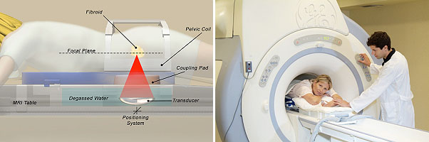
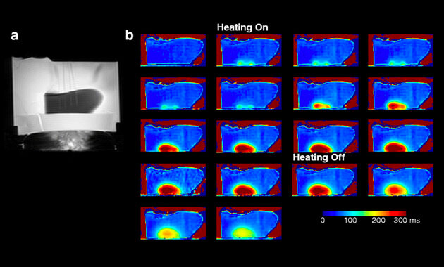
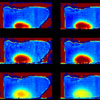
HIFU stands for "High-Intensity Focused Ultrasound." It is also known as MRgFUS (MRI-guided focused ultrasound) and FUS (focused ultrasound). HIFU is an innovative, non-invasive treatment for a wide range of tumors and diseases. HIFU uses an ultrasound transducer, similar to the ones used for diagnostic imaging, but with much higher energy. The transducer focuses sound waves to generate heat at a single point within the body and destroy the target tissue. The tissue can get as hot as 150-200°F in just 20 seconds. This process is repeated as many times as is necessary until the target tissue is destroyed. MRI images are used to plan the treatment and monitor the degree of heating in real time.

What are the benefits of Bone/Musculoskeletal HIFU?

  • Non-invasive non-surgical treatment
  • Lower treatment risks
  • No ionizing radiation
  • Faster recovery time
  • Precision

Clinical Indications

We are currently treating patients with the following indications:

HIFU Clinical Research Trials

HIFU is FDA-approved for treatment of metastatic bone pain. UCSF offers clinical treatments for bone metastases and desmoid tumors which are billed to patient insurance.

We also have two ongoing clinical research trials for patients with osteoid osteomas and undifferentiated pleomorphic sarcomas. For more information regarding these two research trials, please contact our Clinical Research Coordinator, Maya Aslam at [email protected].

HIFU FAQs

How is FUS (Focused Ultrasound) different from standard ultrasound?

Standard ultrasound (i.e. diagnostic ultrasound for imaging) uses low energy ultrasound waves to create images of structures in the body. The energy is not focused on a single spot and does not create high temperatures.

What are the benefits of focused ultrasound for tumor ablation (killing)?

FUS has several attractive features when compared to other methods for non-invasive or minimally invasive tumor ablation such as vascular embolization, radiofrequency ablation, cryotherapy, and targeted radiotherapy. These advantages include:

  • Unlike radiation therapy, the passage of ultrasound energy through intervening tissue has no cumulative effect on that tissue or tissue beyond the focus. Focused ultrasound treatment is extremely precise, with the boundary zone between necrotic treated and viable untreated tissue measuring less than 1 mm. Accordingly, FUS is easily repeatable, unlike radiotherapy where tissue toxicity makes repeated treatment problematic.
  • FUS produces in-depth precise tissue necrosis using an external applicator, with no need to insert an instrument into the target tissue or blood vessels. That is, the procedure is completely incisionless and non-invasive and so carries negligible risks of inducing hemorrhage or infection.
  • Patients treated for bone lesions or soft tissue tumors with FUS are able to return home within 1-2 hours of their procedure, experience rapid and substantial pain relief within a few days, and are often able to reduce or completely stop analgesic use shortly thereafter.
  • FUS requires just a single session of treatment, unlike the multiple sessions required for traditionally fractionated radiotherapy.
  • FUS employs a near-instantaneous delivery of focused energy and does not depend on heat conduction to treat the tumor. Therefore, FUS can be used even in the presence of large adjacent vessels which can act as heat-sinks and limit tumor killing by other modalities such as radiofrequency ablation.
  • The tissue necrosis produced by FUS is immediate and efficacy can be checked by obtaining gadolinium enhanced MR images to show the non-perfused treatment volume at the end of the procedure.

Why not use diagnostic ultrasound to guide and monitor HIFU?

MRI provides two key advantages for guiding focused ultrasound.

First, the ability to rapidly obtain multiplanar and multiparametric images allows for accurate tumor localization, improving FUS targeting.

Second, MR thermometry allows for real-time pixel-by-pixel temperature monitoring during the sonication, so that the extent and degree of tissue heating can be controlled and monitored. Other methods of guiding FUS, for example, the use of endorectal ultrasound for prostate therapy do not provide these advantages of precise tumor localization and treatment monitoring.

Is HIFU safe?

HIFU offers a very favorable safety profile. The most important risk is the risk of a skin burn. In order to minimize this risk, cooling devices and procedures are used to protect the skin during the treatment procedure.

What happens before a HIFU procedure

Several tests and discussions are required before performing an MRgFUS procedure, in order to make sure that only appropriate patients who are likely to benefit from the procedure and who do not have any risk factors that would be a contraindication to performing the procedure are selected. The exact requirements vary with the condition being treated, but at a minimum MRI of the treatment target is required in all cases. It is preferable but not essential for this MRI to be performed at UCSF.

What happens during a HIFU procedure

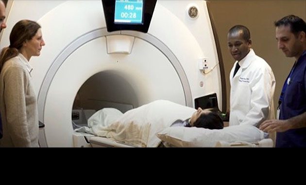
The length of the procedure is variable, depending on what condition is being treated, but most cases last at least 3 hours. In general, the area of treatment is positioned so that the skin overlying the target is centered on the treatment table. The patient receives general anesthesia so that they do not move during the procedure. (They may also receive a nerve block to help decrease post-procedure pain.)

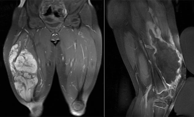
Initial images are obtained to confirm the target is still accessible and appropriate. Then separate pulses (“sonications”) of focused ultrasound energy are applied through the skin using a transducer that is in the table top of the MRI scanner, each lasting about 20 seconds. At the end of the study, an injection of an MRI contrast dye (“gadolinium”) is injected to confirm that the target has been successfully destroyed.

HIFU procedure

What happens after a high intensity focused ultrasound procedure? What are the side effects?

After MRgFUS, patients are allowed to rest comfortably while the effects of any medication given are allowed to wear off. Pain is managed with pain medication. Most patients are able to return to normal activities a few days later. Although, patients who have treatments of bone lesions in the lower extremities may be asked to avoid high impact activities (e.g. running) for up to 4 weeks following the procedure to minimize the risk of bone injury.

Patients who are part of a clinical trial may be asked to fill up questionnaires or other forms in the weeks and months after the procedure.

Clinical Partners

  • Rosanna Wustrack, MD
  • Melissa Zimel, MD
  • Richard O’Donnell, MD
  • Kristopher Jackson, NP
  • Jordan Kaitz, RN
  • Lindsay Lawless, RN
  • Ross Okimoto, MD
  • Victoria Wang, MD, PhD
  • Sorbarikor Piawah, MD
  • Brian Schulte, MD
  • Lawrence Fong, MD

Bone/Musculoskeletal HIFU Team

Associate Professor
Associate Chair, Wellbeing and Professional Climate
Associate Professor In Res.
Professor, & Division Chief, Musculoskeletal Radiology
Clinical Director of MQIR
Director, T32 Program
Professor in Residence
Assistant Professor
Assistant Adjunct Professor
Clinical Research Manager
CLIN RSCH SUPV 2