The Promise of Mid-Field MRI

Magnetic Resonance Imaging (MRI), plays a pivotal role in healthcare, determining treatment paths and eligibility for procedures like hip or knee replacements. Yet, approximately 90% of the global population lacks access to this vital technology, creating barriers to essential care, particularly in rural or remote areas, and even within parts of the United States.
The prospect of a more compact, affordable, and easily installable MRI machine raises an intriguing question: How many lives could be saved, and how much suffering could be alleviated with improved access to this transformative technology?
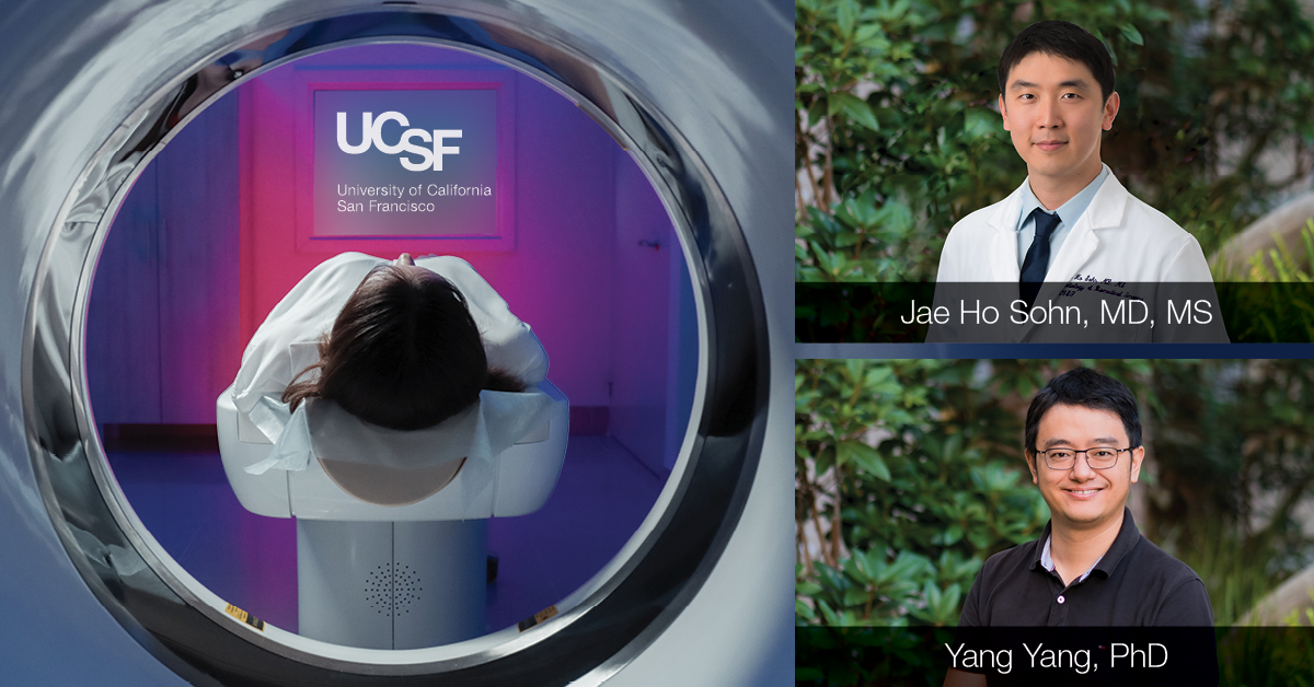
In 2021, UCSF became the first of a handful of healthcare organizations to acquire a mid-field MRI, the Siemens 0.55T Free Max. Since then, UCSF has been conducting research to better understand the promise of these machines and to improve the artificial intelligence associated with them.
“Mid-field MRIs use less space, less power, and can be more accessible,” said Yang Yang, PhD, associate professor of radiology and director of mid-field MRI. Historically, MRI development focused on increasing field strength, reaching up to 7T and beyond. However, the mid-field strength, often overlooked, holds substantial potential for innovation, according to Christopher Hess, MD, PhD, chair of the radiology department. “We have to provide the evidence that it’s an effective screening tool beyond what is currently available,” Hess said.
Mid-field MRI has significantly lower field strengths than conventional MRI, yet there are many reasons to use it. For one, it has the potential to bring the life-saving technology of MRI to more people. Conventional MRIs are expensive, heavy and require special piping to be installed in a room in a hospital. Mid-field MRIs require none of that. In fact, they can be utilized on the trailer of the truck with the potential to bring them out into the community.
“In the future, we might bring the scanner to the patient, enhancing accessibility and potentially revolutionizing routine follow-ups or screenings,” Yang said.
Hess agrees: “When you have a 0.55 T scanner there’s more promise to implement that at scale,” he said. “You’re able to deploy more scanners in the community because it’s less expensive and more accessible.”
“Lower cost and easier installation make mid-field scanners a practical solution for various locations,” Yang said.
Leveraging artificial intelligence, Yang aims to enhance quality and reduce scan times. Preliminary results show promising outcomes, with some scans reduced by 50-73%. “The ideal future is a full stack of AI tools, from acquisition to image reconstruction, enhancement, post-processing, contour drawing, and reporting,” Yang said.
The Clinical Benefits of Mid-Field MRI

Because of the different contrast mechanisms, mid-field MRIs have the potential to screen for some conditions more effectively than current technologies.
Jae Ho Sohn, MD, MS, a cardiothoracic radiologist and assistant professor who is conducting research with Yang, said that patients with lung conditions can benefit from mid-field MRI. “Traditionally, we’ve pushed MRIs to be high field, but in situations like the lungs, lower field is more helpful.”
Mid-field MRI excels in anatomic lung evaluation, offering advantages over traditional CT without radiation exposure. In the lungs, air can cause image degradation, and lower field MRI – in combination with artificial intelligence – can compensate for motion challenges.
“Mid-field MRI is useful for anatomic evaluation of lung parenchyma, offering advantages over traditional CT, involving no radiation and allowing tissue characterization,” Sohn said. "Functional MRI allows us to examine ventilation and perfusion patterns, providing valuable information for characterizing diseases and assessing their severity." Sohn is conducting studies to explore the full clinical potential and limitations of the 0.55T Free Max machine, including correlations with CT for diverse pathologies like cancer, infection, and lung diseases.
Traditionally, since cardiac and lung MRIs are offered separately, a combined cardiac and pulmonary screening protocol that Sohn’s team is developing using mid-field MRI can save a significant amount of time, improving the patient experience. Machine learning will then be used to improve image quality and expedite the scanning process. The team is also developing predictive models to address potential image degradation proactively. This modality offers cross-sectional imaging of the chest without radiation, making it a valuable option for radiation-sensitive populations like pregnant patients.
The wider bore of mid-field MRI, exemplified by the 0.55 Free Max, brings additional benefits. It accommodates patients with claustrophobia or obesity, ensuring accessibility for diverse populations. For individuals with implanted devices like pacemakers, artificial joints, or hips, mid-field MRI becomes a viable option.
Because of the many benefits of mid-field MRI, Yang and Sohn are working to amplify its potential and provide evidence of its success as a viable alternative to conventional MRI through scientific publications, presentations and other channels. “We’re striving to make this transformative technology as widely known as possible,” Yang said.
By Rebecca Wolfson
