

New Contrasts Allow Imaging of Cortical Layers via Myelin Structures
Sometimes discoveries come when a researcher looks out at the edge of their data and says, “Oh, that’s interesting.” Susanne Mueller, MD, recently published “7T MP2RAGE for cortical myelin segmentation: Impact of aging” in PLOS One, in which she shows an imaging technique with great potential beyond its initial conception.

Mueller researches disorders of the brain at UCSF and the San Francisco VA Medical Center. She is particularly interested in the impact of treatment resistant epilepsy on the brain, in particular its potentially deadly consequences. The top mortality risk in epilepsy is known as sudden unexpected death in epilepsy, where in the aftermath of a seizure and an apparent recovery respiration and cardiac function can abruptly stop for reasons that are not yet understood. Mueller’s research showed that the brainstem was involved in these tragedies. However, on a standard 3T clinical MRI scan it is difficult to discern the details of the internal structure of the brain stem that is a relatively small but highly complex structure made up of densely packed and often not well-delineated fiber tracts and small nuclei. Even on a 7T research scanner, a researcher would need a level of highly detailed segmentation that is not available in a traditional MRI. So, Mueller developed a new image segmentation technique that was based on a 7T MR sequence named MP2RAGE that could provide the detail she needed to explore the interactions of epileptic seizures and the brain stem.
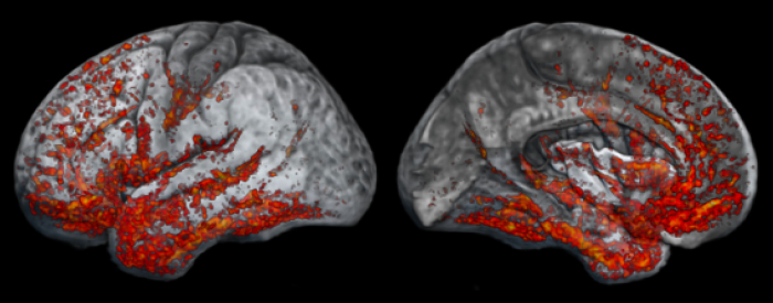
Once I looked at the whole brain rather than just the brain stem, I noticed an intensity gradient in the cortical rim in one of the MP2RAGE contrasts that seemed to follow the cortical myelination pattern described in histological preparations that I hadn’t seen before. That’s when I thought, ‘Oh, this is very useful!’ That’s how this project came to be.
Susanne Mueller, MD, Professor, Radiology
Cortical grey matter (GM) is separated into cortical layers that cannot be distinguished in traditional imaging. However, the cortex is also divided into heavily myelinated and sparsely myelinated portions, which have a high correspondence to the cortical layers. Mueller discovered that the MP2RAGE-derived contrasts returned results which could clearly separate them by linking the different degrees of myelination to the cortical layers. These cortical layers, each less than half a millimeter thick, have different functions and are connected differently to other parts of the brain.

Multiple diseases of the brain manifest differently by layer, making accurate imaging of these delicate structures of great value. For example, Alzheimer’s disease manifests volume loss at the top or superior cortical layers, while chronic traumatic brain encephalopathy (CTE), which can also cause dementia, manifests at the lower layers. A scan that can read the rates of volume loss at the different layers of the cortex may more easily differentiate between the two.
Mueller’s new segmentation techniques have an advantage over prior methods proposed for imaging cortical layers: they do not require special imaging with long durations. Though Mueller developed her methods on 7T scanners, she found that it is possible to acquire the same type of sequence on a standard 3T imaging suite at a slightly lower resolution but still of usable quality. When adapting the strategy to 3T, implementing a few extra measures in image acquisition compensates for the lower resolution and raises the scan time from 8 minutes to 12 minutes.
 This novel imaging technique for cortical myelin has the potential to benefit patients with epilepsy. One goal for epilepsy treatment is to pinpoint and localize the relevant areas of the brain to stop seizures without impeding critical functions in adjacent parts of the brain. By using this cortical segmentation technique, clinicians might be able to more precisely target that region within the brain to provide relief to patients with medication-resistant focal epilepsy.
Mueller sees many avenues forward from these unexpected 7T imaging insights into brain structure, “Right now, I would apply this technique to every disease I could get my hands on just to figure out what it can do: schizophrenia, multiple sclerosis, as well as autism and developmental disorders. There is great potential for research that images the ongoing myelination of developing brains. We’re just scratching the surface of MP2RAGE-derived contrasts at 7T, and I look forward to seeing the many potential avenues this research can take.”