
Overall, many people are unaware of the importance of osteoporosis screening and preventive measures that can be taken. Regular screening can diagnose osteoporosis and other bone problems early. From there, they can be managed and even curbed with prescription medication and lifestyle modifications. At the UC San Francisco Department of Radiology and Biomedical Imaging, we support recommendations by the National Osteoporosis Foundation that women age 65 and older and men age 70 and older undergo regular bone density screening tests. However, we also want to make patients and referring physicians aware of risk factors for early osteoporosis screening that can reduce the screening age to 40 years old for some candidates.
Those who are at high risk include:
- People with a family history of osteoporosis
- Those of Asian or Northern European descent
- Men and women over the age of 50 who suffer fractures
- Young, competitive female athletes who exercise too much and eat too little, resulting in amenorrhea and risk for low bone mass and fractures
- People with low BMI
- People who have suffered from an eating disorder
There are also lifestyle risk factors, including:
- Smoking
- High alcohol intake (three drinks per day or more over a long period of time), or the intake of other drugs
- A diet with insufficient calcium intake
- Vitamin D deficiency due to lack of sun exposure or other factors
- People who under-exercise
Additional risk factors related to health include:
- Rheumatoid arthritis
- Chronic kidney disease
- Early menopause (from natural causes or surgery)
- History of hormone treatment for prostate or breast cancer
- Taking corticosteroids (prednisone, methylprednisolone) every day for three months or more
- Sudden, significant loss in height, which can put men and women at risk for fractures
Assessing bone health is very important. According to the International Osteoporosis Foundation, there are 44 million people in the U.S. with either osteoporosis or low bone mass. This number represents 55 percent of those age 50 and older. Osteoporosis weakens bones, leaving both men and women subject to hospitalization due to fractures. At UCSF Radiology, Thomas Link, MD, PhD, has focused his research on degenerative diseases of the musculoskeletal system such as osteoporosis and osteoarthritis. His goal is to develop imaging tools which allow better assessment of the risk of an individual patient to develop debilitating and painful degenerative joint disease, leading to therapy and lifestyle modifications which can prevent the disease from either occurring or progressing.
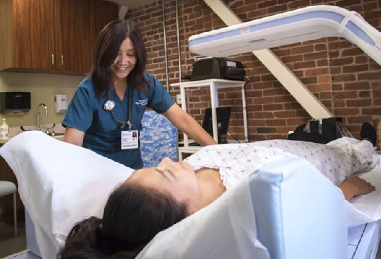
Dual-energy X-ray absorptiometry (DXA or DEXA) bone density scanning is used to determine bone density and strength. The lower the bone density, the greater the risk for fracture. It is the most accurate method available for the diagnosis of osteoporosis, as well as an accurate estimator of fracture risk. It's also painless, takes about 10 minutes and uses a low amount of radiation (about 10 percent of a normal chest X-ray). An alternative to DXA is quantitative computed tomography (QCT). QCT exams can measure bone density in patients who are very small or large, patients with degenerative disease of the back, and patients with prostate cancer who are being treated with androgen deprivation therapy.
Both DXA and QCT are available at a number of UCSF Radiology locations, including Montgomery Street, Mission Bay and Mount Zion. To learn more or schedule a DXA exam, speak to your referring physician or call (415) 353-3900.