Combining Advanced Imaging Techniques with Loading Biomechanics to Slow, Prevent and Treat Osteoarthritis
Osteoarthritis is a common disease with limited treatment options at this time. In his lab, Richard Souza, PhD, PT, and his team are working on lower extremities biomechanics research, looking at people with diseases of the lower extremity, such as hip and knee osteoarthritis (OA), and how they move. The researchers use 3D motion analysis to carefully quantify the movement patterns of patients when they are walking or climbing stairs or rising from a chair. The researchers also look carefully at patients’ joints using quantitative MRI techniques that have been developed by UCSF radiologists. By following these people over time, Dr. Souza’s team can determine which patients are getting worse in their disease and which movement patterns are predictive of disease progression.
As both a professor in the UC San Francisco Department of Radiology and Biomedical Imaging and vice chair of research in the Department of Physical Therapy and Rehabilitation Science, Dr. Souza knows that multi-disciplinary collaboration is very important. In its studies, his team uses specialized image pulse sequences developed by imaging scientists to evaluate the health of a joint. The researchers then collaborate closely with radiologists at UCSF so that they can look at the lesions to the cartilage, changes in the bone marrow edema, and at other structures that are important in the OA disease process.
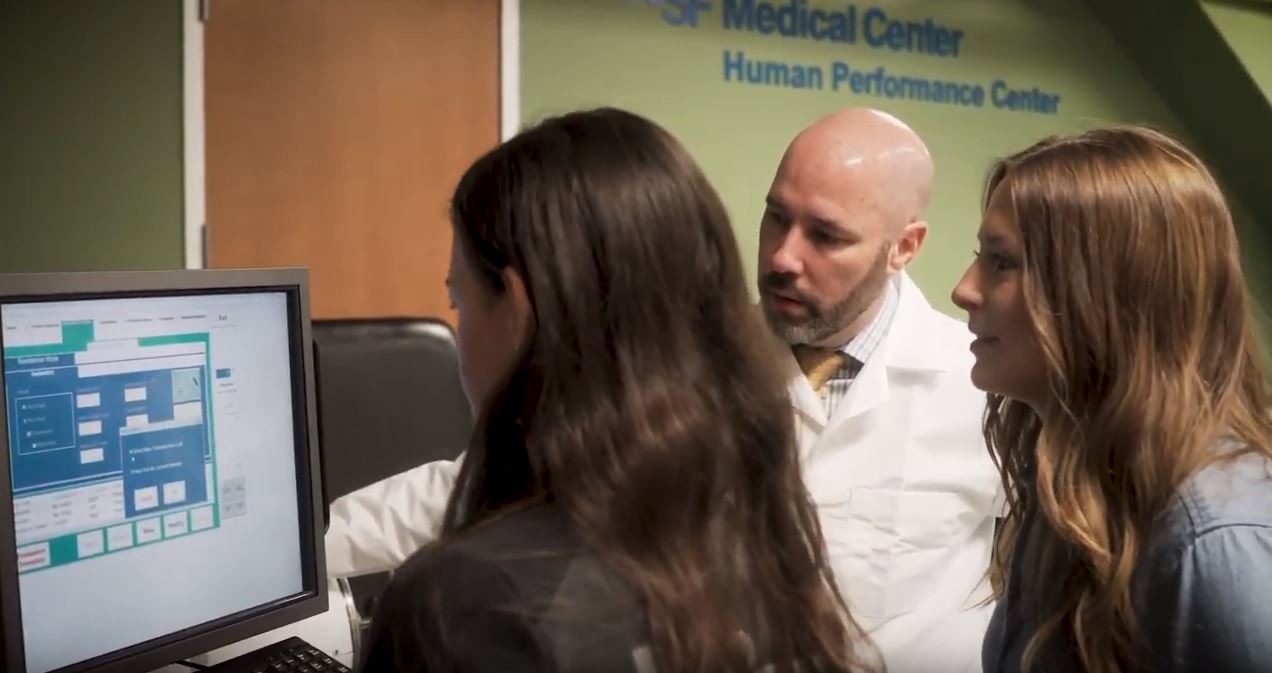
In the accompanying video, hear more from Dr. Souza and see the work his team is doing to change loading profiles and intervene in the OA disease process.
Visit the Musculoskeletal RIG web page to learn more about the Musculoskeletal Research Interest Group (RIG) and how these scientists are exploring the structures that support the human body, their role in health, and how to prevent and heal musculoskeletal damage.