

Celebrating Our Sonographers: Medical Ultrasound Awareness Month
This Medical Ultrasound Awareness Month, we’re proud to recognize the extraordinary skill and expertise of our sonographers. Most known for letting us sneak a peek at babies during pregnancy, ultrasound is also used to visualize internal organs, provide real-time biopsy guidance for a variety of medical conditions, and more.
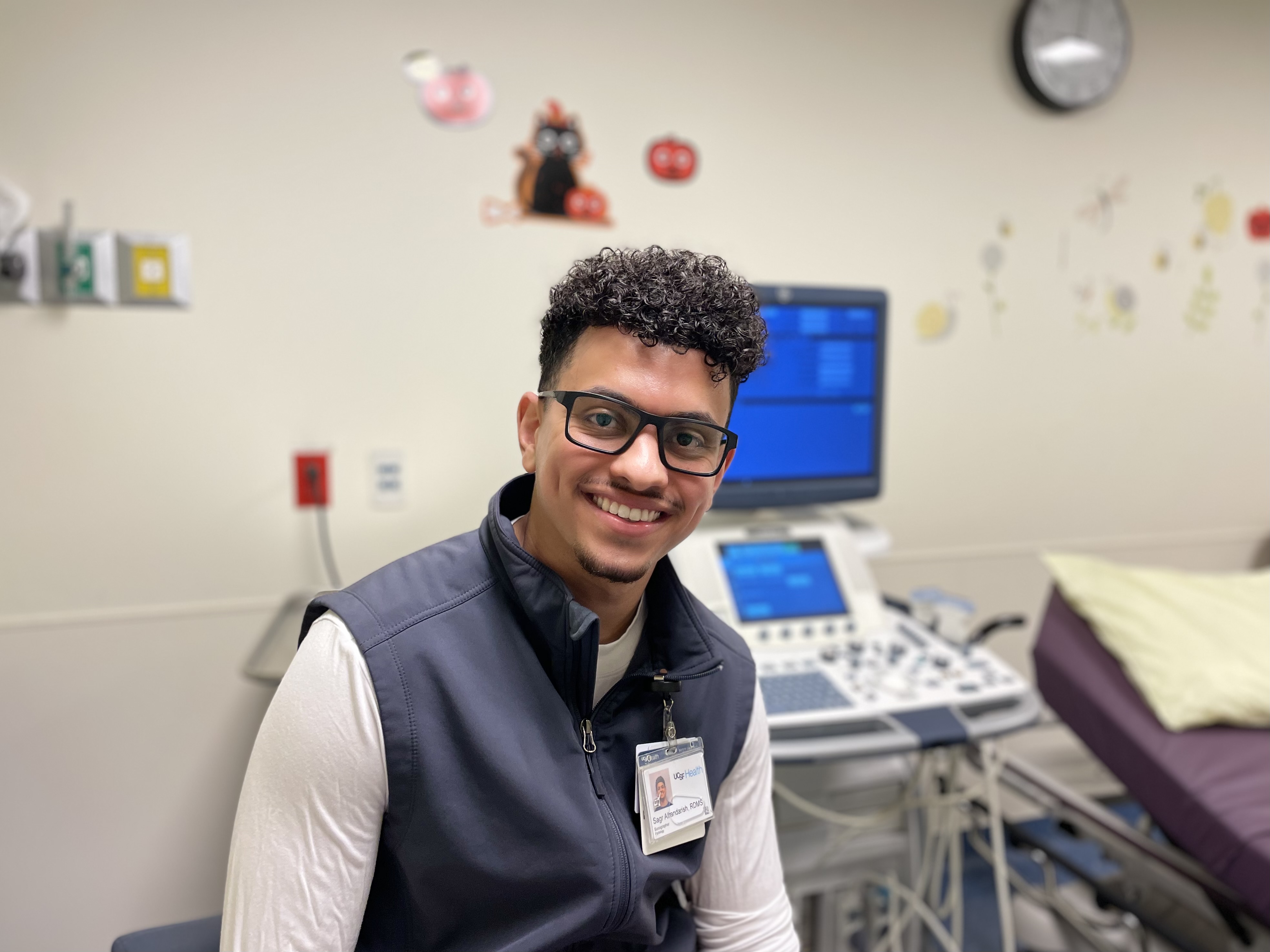
“Memorable moments are made each shift,” says Sagr Alhandarish, an ultrasound sonographer at Mission Bay. “One in particular, is a two-year-old patient I first met over a year ago when she received a liver transplant. She is now thriving and has the most adorable verbiage during her follow-up studies.”

“As a new grad, having the opportunity to work for UCSF has been an absolute dream come true,” says Mikaela Tigno, an ultrasound technologist who helped open Mission Bay’s Bayfront Medical Building in August. “Working alongside and learning from my fellow sonographers in this new environment has been such an amazing experience.”
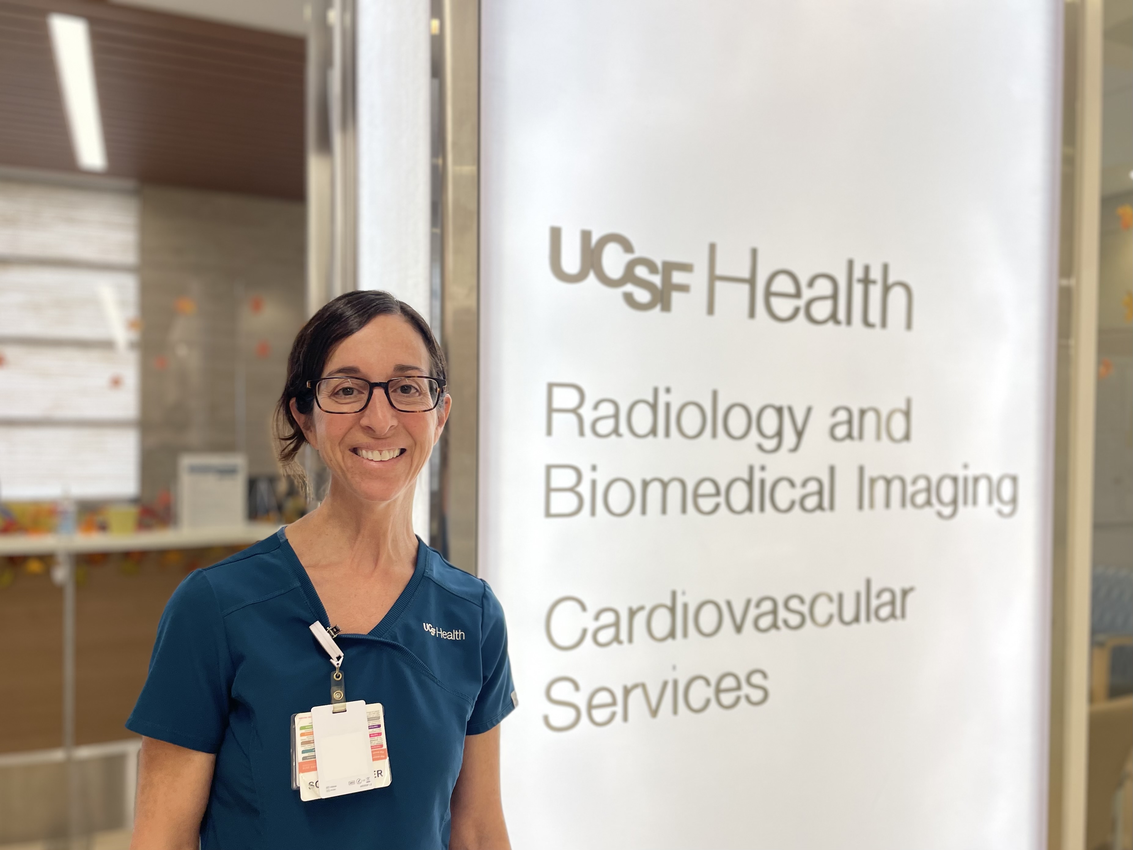
“What I enjoy most about being a sonographer is the level of human connection involved in my job and the valuable diagnostic role I play in a patient's medical care,” says Jane Glover, RDMS, RVT, a principal ultrasound technologist with more than 22 years of experience. “The fact that the images I take offer a clue—a piece of the puzzle in making a diagnosis that can help direct a patient's care—is truly rewarding.” Jane also highlighted the vital role ultrasound imaging plays before and after organ transplant procedures – one of her personal favorites to scan.

“Ultrasound is so much more than just looking at babies; we look at transplants, muscles and nerves, arteries and veins,” adds Principal Sonographer Angela Lee, RDMS, RVT, RMSKS, who describes her role as a sonographer as incredibly rewarding. “Sometimes we are the first people to see a patient's cancer or birth defect.”
Career Advice
“Shadow a sonographer for a day or two and see what their workday is like,” Jane advises those interested in a career in sonography. She recommends ultrasound programs recognized by the Commission on Accreditation of Allied Health Education Programs (CAAHEP), which enable graduates to take exams and earn credentials.
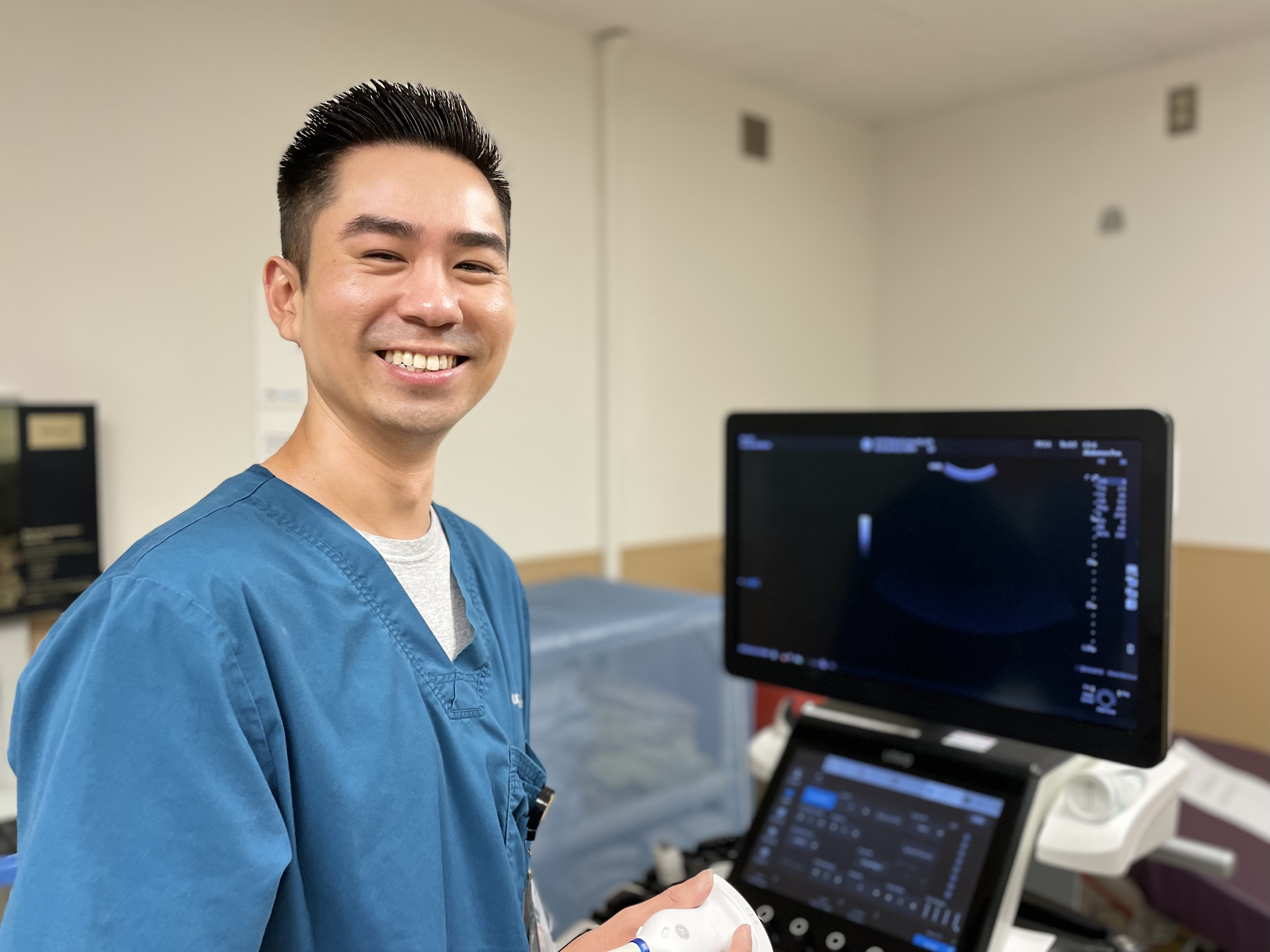
“There will be highs and lows when it comes to learning how to scan but it is all part of the learning process!” emphasizes Senior Ultrasound Technologist Brent Yim, RDMS, reflecting on his own career in ultrasound. “Always remain humble and remember what it was like starting out your career.”
Ever-Evolving Technology
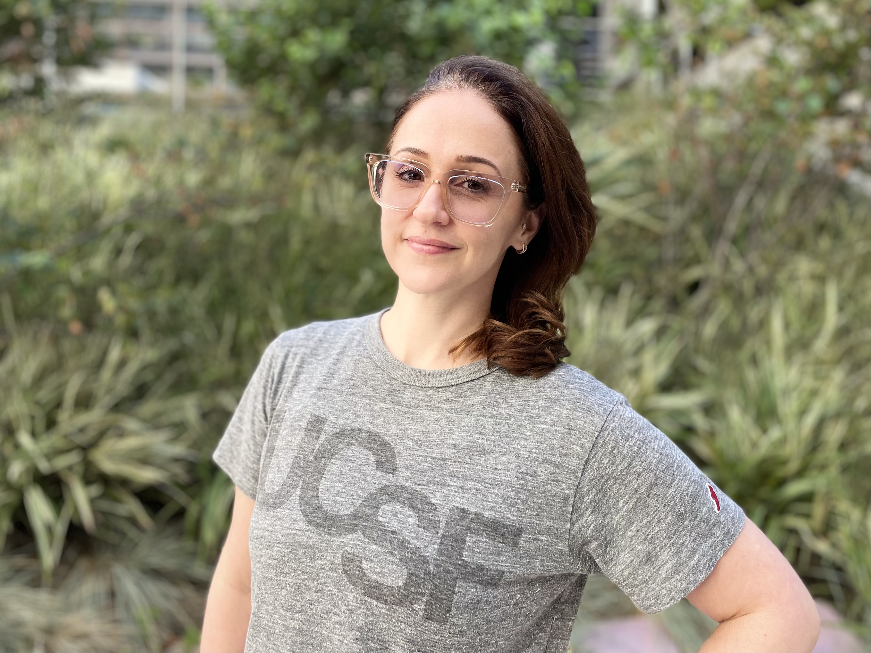
"I love that my career offers continual learning and growth, allowing me to adapt to challenges and the ever-evolving technology," says Principal Sonographer Lisa Valentinsson, RDMS, RVT. It’s both meaningful and rewarding to her to be a sonographer and provide care for patients, and she enjoys sharing her knowledge with sonography students at UCSF.

“Be prepared for both technical challenges and emotional moments,” says Irene Sun, who notes she learns every day and agrees that ultrasound is continuously evolving with the latest technology advancements. She highlights how impactful ultrasound imaging can be for early detection in a patient’s healthcare journey, and shares: “During a routine ultrasound, I discovered an early-stage tumor that helped save a patient's life.”
At UCSF Radiology, we appreciate the compassion and dedication of our talented sonographers, and we extend our thanks for their contributions to exceptional patient care.
3 Tips for Patients
- Ultrasound is considered a very safe imaging modality that uses sound waves instead of radiation to produce images of internal body structures.
- Ultrasounds are generally painless exams that involve the use of gel and a handheld transducer
- Most people associate ultrasound with visualization of the fetus during pregnancy. Ultrasound can do that and so much more, including diagnostic visualization of organs (liver, kidneys, thyroid, etc.), real-time guidance for biopsies, and ablative procedures using high-intensity (HIFU) or low-intensity (LIFU) focused ultrasound.
-- Jane Glover, Principal Ultrasound Technologist, RDMS, RVT, Parnassus Heights.