
With doctor specialists dedicated to ultrasound, UCSF’s sonographic expertise is distinct from that of other medical centers. Because we perform a very high volume of ultrasounds and are responsible for a large number of abnormal scans, our level of expertise in using ultrasound for diagnoses and treatment recommendations exceeds that of most medical centers in the nation. We have more than 100 years of dedicated ultrasound experience.
The UCSF Ultrasound Subspecialty has advanced experience and specialization in:
Fetal Imaging
Indepth Experience - Normal and High Risk
UCSF’s ultrasound specialists are experts in fetal development and perform approximately 3,500 fetal ultrasound studies per year. The ultrasound subspecialty has been instrumental in developing and supporting a growing Fetal Treatment Center at UCSF. We perform all of the high-risk obstetrical sonograms of patients referred to UCSF for potential fetal therapy, and in this role we consult and collaborate with the perinatologists, geneticists and pediatric/fetal surgeons.
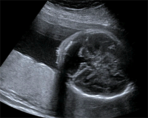
Cardiac teratoma and pericardial effusion.
Our physicians are experts in the diagnosis of fetal anomalies including:
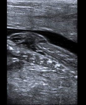
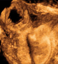
Myelomeningocele high resolution ultrasound.
- Twin syndromes such as Twin-Twin Transfusion Syndrome (TTTS) and Twin-Reversed Arterial Perfusion syndrome (TRAP).
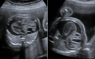
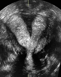
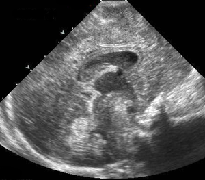
- Fetal brain and spine anomalies including Spina Bifida, Fetal Ventriculomegaly, Dandy-Walker Malformation, and Agenesis of the Corpus Callosum.
Dandy Walker Malformation.
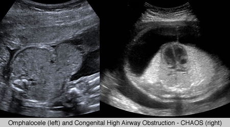
UCSF doctors in ultrasound are also expert in fetal thoracic and abdominal pathologies, including gastroschisis, omphalocele, lung sequestration, and cystic adenomatoid malformation (congenital pulmonary airway malformation) and congenital high airway obstruction (CHAOS). We assist in fetal operations for these anomalies by providing guidance for radiofrequency ablation, laser ablation for Twin-Twin Transfusion Syndrome, fetal blood transfusions, fetal lung mass resections, tracheal balloon placement and fetal surgery for Spina Bifida. (Open myelomeningocele)
Imaging for Organ Transplantation
The high volume of transplant patients at UCSF requires that our ultrasound physicians have special expertise in complex abdominal pre- and post-operative transplant sonography. In addition, UCSF’s ultrasound physicians guide or perform more than 800 organ transplant biopsies per year.
Gynecologic Imaging
UCSF‘s ultrasound subspecialty uses state-of-the-art, multimodality approach to gynecologic imaging processes (including endometrial evaluation with and without saline infusion) to diagnose ovarian and uterine pathology as well as complications of early pregnancy, including ectopic pregnancy. We contribute to improved patient outcomes by using image guidance for methotrexate injections and bedside sonography in the operating room in challenging gynecologic surgeries.
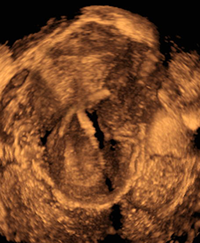
Septated uterus, 3D image.
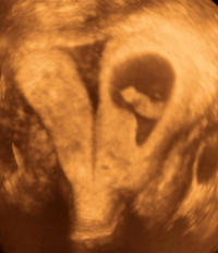
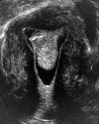
Septated uterus, pregnancy in left horn, 3D image.
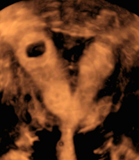
Septated uterus, pregnancy in right horn, 3D image.
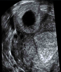
Cornual insterstitial ectopic pregnancy, 3D image.
Cornual insterstitial ectopic pregnancy, 3D image.
Malpositioned IUD, 3D image.
Endometrial polyp on sonohysterogram, 3D image.
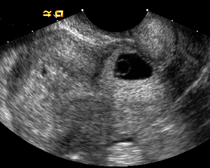
C-section scar ectopic pregnancy.
Intra-Operative Ultrasound
UCSF utilizes intra-operative ultrasound, which is an ultrasound technology for both diagnosis and treatment that is used during surgical procedures performed in the operating room. Because of the portability and high resolution of intra-operative ultrasound, UCSF surgeons find it to be a particularly useful tool for making real-time decisions during a surgery, for guiding biopsies in adults and children, and for guidance of intra-operative fine needle or ablation probes.
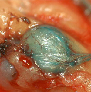
Speciman of metastatic lymph node after blue dye injection.
Intra-operative ultrasound has also emerged as an important tool:
- In accurately staging metastatic cancer such as in liver or colorectal disease,
- For intra-operative injections of “blue dye” into small metastatic lymph nodes (see image above),
- To assist transplant or liver surgeons in assuring that the vascular anastamoses are not blocked before the patient leaves the operating room, and
- To aid in delicate fetal surgical therapies such as radiofrequency ablation or fetal thoracic fluid drainages.
Reference:
Harari A, Sippel RS, Goldstein R, Aziz S, Shen W, Gosnell J, Duh QY, Clark OH: Successful Localization of Recurrent Thyroid Cancer in Reoperative Neck Surgery Using Ultrasound-Guided Methylene Blue Dye Injection. J. Am. Coll. Surg. 2012; Oct 215(4):555-561.
Pediatric Imaging
Dramatic advances in imaging technologies are improving the ability of pediatric radiologists to diagnose complex cases in children. For all imaging of children at UCSF we pay special attention to ensuring that protocols are pediatric specific and that all imaging studies are interpreted by radiologists who are specially trained in pediatric anatomy and pathology. UCSF ascribes to the policy of “imaging gently“[1] in the imaging of children, and the use of ultrasound is an important tool in achieving that goal.
UCSF’s ultrasound subspecialty has world-renowned experts in prenatal ultrasound imaging as well as for children and babies. Our expert staff includes specialists in prenatal imaging of congenital anomalies and fetal therapy.
Pediatric cranial sonogram.
[1] Image Gently: The American College of Radiology has released new guidelines on the subject of imaging children and babies, and the Alliance for Radiation Safety in Pediatric Imaging, a consortium of professional societies concerned about the issue, has launched an “Image Gently” campaign to promote safe practices; UCSF is an active participant in that campaign.
Further attesting to our expertise in fetal imaging, UCSF is one of the three centers that performed the prenatal imaging for a recently published New England Journal of Medicine article on prenatal surgery for myelomeningocele.