
UCSF's Ashish Raj, PhD, Explores Abnormal Brain Activity in Alzheimer's Disease
A study from the UCSF Department of Radiology and Biomedical Imaging investigates the relationship between aberrant neuronal circuit mechanisms and functional abnormalities observed in Alzheimer's disease (AD) using magnetoencephalography (MEG) imaging. AD is a multifaceted neurodegenerative condition that gradually erodes cognitive function, presenting significant challenges for patients and their families. Despite extensive research, there is still a lot we don't understand about its underlying mechanisms.
 "Impaired long-range excitatory time scale predicts abnormal neural oscillations and cognitive deficits in Alzheimer's disease," published in Alzheimer's Research & Therapy, was lead-authored by Parul Verma, MD, and overseen by Ashish Raj, PhD, director of the Brain Networks Laboratory at UCSF. It sheds new light on how AD affects the brain's functional activity patterns. The research team used magneto-encephalography (MEG) to look at brain activity in people with AD and compared it to people without the disease. They wanted to see how brain signals were different in AD and how these differences might relate to thinking and memory problems. Analyzing MEG data from AD patients and age-matched controls, the researchers aimed to reveal the overall alterations in neural mechanisms associated with the spatial and spectral electrophysiological patterns in AD.
Neural synchronization, the collective and concerted activity of billions of neurons in the brain, is pivotal for cognitive function. In AD, disruptions in both local and long-range synchrony suggest a possible connection between abnormal synchrony and cognitive decline. In order to understand the neural processes that may cause this disruption, researchers employed a new in-house mathematical model they have recently developed, called the Spectral Graph Model (SGM). "SGM is a biophysically realistic and analytical model. Its analytical nature makes model computations straightforward and efficient," notes Verma. "Additionally, the parameters inferred from this model have biophysical interpretability, and therefore can be readily used in understanding the underlying mechanisms of a disease." 
"In this study, we determined local and long-range neuronal parameters of this computational model of brain activity that can account for abnormal neurophysiological activity in AD observed in high spatio-temporal resolution MEG imaging," said Raj.
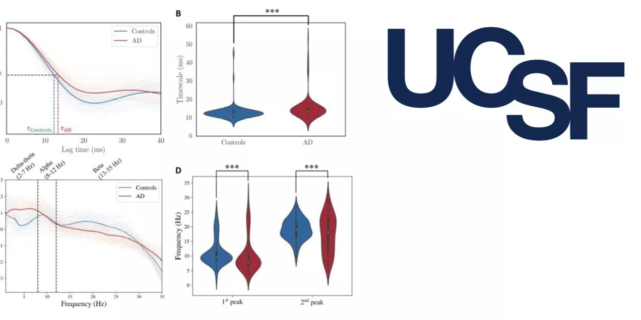
This study identified three significant findings. First, AD patients exhibited notably increased long-range excitatory neuronal time scales compared to controls, suggesting that the overall speed of signal transmission in the brain slows down due to disease. Second, this heightened long-range time scale correlated with more severe deficits in the subject's cognitive performance, indicating its potential as a predictive marker for cognitive decline in AD. Lastly, the study suggests that the long-range excitatory time scale acts as a fundamental factor underlying altered neuronal activity in AD, connecting the dots between abnormal brain signals and the big-picture brain problems seen in AD.
These findings hold considerable implications for understanding and managing AD. By unraveling the neural mechanisms behind abnormal synchrony patterns, researchers may pave the way for novel diagnostic and therapeutic approaches. Identifying the long-range excitatory time scale as a potential biomarker for cognitive decline could aid in early detection and intervention strategies.
By elucidating the role of the long-range excitatory time scale in shaping neuronal activity, this study offers valuable insights into the disease's pathophysiology. Ultimately, these findings advance our understanding of AD and may facilitate the development of more effective therapeutic interventions in the future.
Led by Raj, the team is now embarking on a similar ambitious study of MEG patterns in Parkinson's disease (PD), a neurodegenerative disease that mainly affects motor and gait functions. They are analyzing brain activity data from Parkinson's patients from the OMEGA, a public multinational study. They will apply the same SGM model and assess whether the elongation of long-range time constant can explain the altered MEG patterns in PD, as it appears to do in AD.
In addition to Raj and Verma, the research team included Kamalini Ranasinghe, MBBS, PhD, Janani Prasad, Chang Cai, PhD, Xihe Xie, Hannah Lerner, Danielle Mizuiri, Bruce Miller, MD, Katherine Rankin, PhD, Keith Vossel, MD, Steven Cheung, MD, and Srikantan Nagarajan, PhD.