Study Results from the UCSF Ci2 Suggest Deep Learning Methods Can Help Grade ACL Injuries

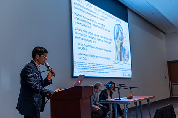
Injuries to the anterior cruciate ligament (ACL) are very common, and ACL injuries increase the risk of developing post-traumatic knee osteoarthritis and total knee replacement (TKR). At present, Magnetic Resonance Imaging (MRI) is the most effective imaging modality for distinguishing structural properties of the ACL in relation to adjacent musculoskeletal structures. Several multi-grading scoring systems have been developed to standardize reporting of knee joint abnormalities using MRI including the Whole-Organ Magnetic Resonance Imaging Scale (WORMS) and the Anterior Cruciate Ligament OsteoArthritis Score (ACLOAS). However, both of these grading metrics are susceptible to inter-rater variability.
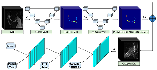
Deep learning methods have recently shown potential to serve as an aid for clinicians with limited time or experience in osteoarthritis grading of the knee menisci and cartilage. Recently a team of scientists from the UCSF Center for Intelligent Imaging (ci2) evaluated the diagnostic utility of two convolutional neural networks (CNNs) for severity staging of anterior cruciate ligament (ACL) injuries. “Previous studies have developed binary classifiers to distinguish fully torn ACLs from intact ACLs,” said Nikan Namiri, medical student at UCSF School of Medicine and corresponding author. “And our study is the first to take deep learning a step further to help classify a broader spectrum of injury, which may be more useful in the clinical setting.”

This retrospective analysis was conducted on 1243 knee MR images (1008 intact, 18 partially torn, 77 fully torn, and 140 reconstructed ACLs) from 224 patients. At the conclusion of their research, scientists determined that 2D and 3D CNNs applied to ACL lesion classification had high sensitivity and specificity, suggesting that these networks could be used to help grade ACL injuries by non-experts. “We studied differences between 2D and 3D convolutional neural networks,” said Nikan Namiri. “Both classifiers demonstrated high performance metrics and there was no substantial difference in using one over the other.”
Overall, this deep learning pipeline could lend towards standardization and generalizability in assessing ACL lesions for clinicians with limited time or those with limited experience reading knee MRIs. The study, with full methodology and results, was published in Radiology: Artificial Intelligence. “The volume of medical imaging has been rapidly increasing and radiologists are struggling to keep up with the sheer number of images,” said Sharmila Majumdar, PhD, professor and vice chair in the UCSF Department of Radiology and Biomedical Imaging. “Models such as this will have a tremendous impact on standardizing, and reducing evaluation time for sports medicine and musculoskeletal imaging"
The team of scientists who worked on this study also included Thomas Link, MD, PhD, and Valentina Pedoia, PhD, faculty in the UC San Francisco Department of Radiology and Biomedical Imaging, along with Bruno Astuto, PhD, assistant researcher, UCSF Ci2; Io Flament, machine learning specialist; Rutwik Shah, postdoctoral scholar; Radhika Tibrewala, assistant specialist and Francesco Caliva, PhD postdoctoral scholar.
The UCSF Ci2 was founded in fall 2019 as a hub for the multidisciplinary development of AI in imaging to meet unmet clinical needs and provide a platform to measure impact and outcomes of this technology.
