
Dual-energy CT (DECT) is not new, in fact, says Benjamin Yeh, MD, it has been around just as long as computed tomography (CT) has. What is new is that CT scanners can capture DECT quickly and with good image quality. The benefits are mostly that it improves confidence and specificity for diagnoses. Like regular CT imaging, DECT provides 3-dimensional information about the size and shape of lesions, along with a fourth dimension in the form of X-ray density measured in grayscale Hounsfield Units (HU). Profoundly, DECT adds a fifth dimension of X-ray density across high-and low-energy spectra.
"With DECT, structures with different atomic make-up, such as iodine, calcium and dense blood or metal, are vividly differentiated," said Dr. Yeh at the RSNA 2019 Annual meeting. "Even when objects have identical HU on a regular CT, with DECT different materials appear like different colors."
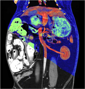
The benefits of these different colors, aka "color vision," makes DECT a transformative technology in daily clinical practice. Dr. Yeh explains that radiologists routinely encounter ambiguous lesions at CT in their daily practice. Since many types of materials may have similar HU values, it is hard to tell whether a lesion is enhancing, calcified or hemorrhagic. DECT shows radiologists what the underlying atoms are and readily differentiates among such entities.
Another benefit of DECT is that it affects that way contrast material is used. With DECT, we get diagnostic-quality images with lower contrast material doses, as are often needed in patients with renal insufficiency or poor IV access, Dr. Yeh points out. DECT derives simulated low-energy images where the HU values of contrast enhancement may be doubled or tripled so imagers can better see hypovascular or hypervascular lesions.can also simulate what the CT scan would look like if the IV contrast had not been given at all. Additionally, Dr. Yeh says that DECT can help to reveal many hard-to-see lesions such as CT-isodense gallstones and bone contusions.
Dr. Yeh is a professor in the Abdominal Imaging Section in the UC San Francisco Department of Radiology and Biomedical Imaging. He is also principal investigator (PI) of the Contrast Material and CT Translational Research Lab. The lab's translational research group focuses on the exploration and development of novel CT techniques for the diagnosis of clinical disease.
One of their projects is to develop vendor-agnostic software to streamline dual energy image processing, improve material separation capability, allow quantitative comparison of images from all CT scanners, and allow double contrast-enhanced DECT to give richly detailed co-registered images of complex abdominal anatomy for millions of Americans at a low radiation dose and major cost savings to the health care system.
Other ongoing projects include the development and testing of novel contrast materials, bowel imaging, fibrosis imaging, assessment of contrast material distribution in healthy and diseased tissues and oncologic imaging. One new contrast agent invented by the lab at UCSF is in early clinical trials, and is hoped to improve lesion detection at abdominal CT. Their research group works closely with the abdominal imaging subspecialty of UCSF Radiology and includes the former Center for Pharmaceutical and Molecular Imaging.
Learn more about their work by visiting the lab's web page.