Cardiothoracic Imaging
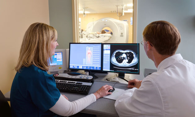
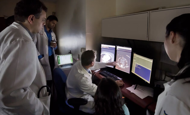
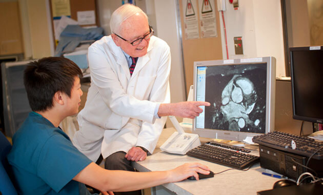
The Division of Cardiothoracic Imaging at UCSF Radiology is dedicated to safely performing the most current clinical imaging exams of both the respiratory and cardiovascular systems using advanced imaging modalities, such as detailed CTA and CT exams.
Why choose Cardiothoracic Imaging at UCSF
- Accurate image interpretation by internationally renowned experts in cardiothoracic imaging
- Optimized CT imaging protocols with as low as reasonably achievable (ALARA) radiation while maintaining excellent image quality
- Optimized MR imaging protocols that achieve excellent image quality while reducing scan duration
- High-volume CT-guided chest procedure team (>3000 cases in the past decade)
- Clinical integration of cutting edge research work in partnership with clinical teams and industry
Who we serve
- Patients suffering from complications caused by cancers/tumors
- Patients who are at risk for lung cancer
- People diagnosed with cancer
- Adult and pediatric cardiac patients
Conditions we frequently address
Cardiac
- Coronary artery disease
- Ischemic Cardiomyopathy
- Non-ischemic cardiomyopathy (including but not limited to myocarditis, amyloidosis, sarcoidosis, arrhythmogenic right ventricular dysplasia, hypertrophic cardiomyopathy, and dilated cardiomyopathy)
- Valvular diseases and transcatheter cardiac valve replacement preparations
- Complex Congenital heart disease
- Complex post-surgical follow up cases in the heart, aorta, and great vessels
- Cardiac transplant follow up
Pulmonary
- Lung cancer (primary lung cancers and pulmonary metastasis)
- Pulmonary embolism
- Interstitial lung diseases (Idiopathic Pulmonary Fibrosis, Sarcoidosis, Hypersensitivity Pneumonitis, Cystic Lung Disease)
- Lung transplantation (pre-transplant and post-surgery follow up)
- Infectious diseases of the thorax (including immuno-compromised cases)
- Smoking related lung diseases
- Airways diseases
- Mediastinal diseases
- Complex post-surgical follow up cases in the lungs, esophagus, and mediastinum
- MRI evaluation of the lungs (investigational UTE sequences)
- Vascular disease of the aorta and pulmonary vessels
Cardiothoracic Imaging Services
- Chest X-rays
- Rib X-rays
- Routine chest CT
- Lung cancer screening CT
- High resolution CT (HRCT) of the lungs
- CT angiography (CTA) of the pulmonary arteries and aorta
- Coronary Calcium Score CT
- Coronary CTA
- Fractional Flow Reserve (FFR) - Coronary CT
- Transcatheter Valve Replacement CTs (including aortic, mitral, pulmonary (Harmony), and tricuspid valves)
- Congenital Heart CT
- Ventricular Scar Mapping CT
- Left Atrial Mapping CT (in prep for cardiac ablation)
- Cardiovascular MRI
- Thoracic Vascular MRA
- Left Atrial Mapping MRA (in prep for cardiac ablation)
- Ventricular Scar Mapping MRI
- Congenital Heart MRI
- Thoracic MR angiography
- UTE Lung MRI (investigational)
- PET-CT (chest focus)
- SPECT myocardial perfusion imaging (by nuclear medicine)
- Cardiac PET (by nuclear medicine)
- Cardiac Stress Testing PET (by nuclear medicine)
- Virtual Bronchoscopy
- Percutaneous CT guided chest biopsies
- Percutaneous CT guided gold fiducial marker placement (in prep for radiation therapy)
- Percutaneous CT guided indocyanine green localizer injection (in prep for surgery)
- Percutaneous CT guided medication injection (for investigational therapy)
Who we partner with
- Patients and their families
- Researchers from our own and other institutions
- Donors and other visionaries committed to improving the lives of others
- Referring colleagues in the fields of Cardiology, Thoracic Surgery, Pulmonary Medicine, Oncology, Infectious Disease and Primary Care/Internal Medicine.
Who we are
- Faculty members
- Cardiothoracic Imaging Fellows
- Postdoctoral fellows
- Research staff
- Medical and graduate students

