UCSF’s Cutting-Edge Use of Fetal MRI: Diagnosing Complex Abnormalities in Utero
 In the United States, a baby is born with a birth defect every 4-½ minutes. That means nearly 120,000 children are affected by birth defects each year, according to the Centers for Disease Control and Prevention. While many birth defects may be managed after the child is born, an increasing number of conditions can be corrected before birth to prevent devastating consequences. These early interventions – such as fetal magnetic resonance imaging (or fetal MRI) – can help babies to survive and thrive.
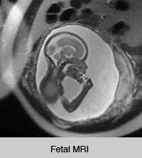
For women who are expecting, ultrasound plays an important role in monitoring fetal development throughout the pregnancy. The imaging technique, however, has its limitations. Specifically, ultrasound’s capabilities to detect certain abnormalities, such as in the brain or spine, are limited. Clinicians at UC San Francisco have adopted the use of fetal MRI to better detect these abnormalities during pregnancy.
There are several benefits to using fetal MRI for detection and treatment of birth defects. When abnormalities are spotted on ultrasound but are not clearly defined or if a fetus has significant risk for an abnormality, fetal MRI represents a practical choice for clinicians. The technique can show physicians and researchers how various abnormalities correlate with childhood development. Further, fetal MRI enables abnormalities to be classified more thoroughly, which can then give parents a more accurate picture of what they can expect for their child from a neurological and developmental standpoint.
UCSF is a world leader in diagnosing and treating birth defects before delivery. The neuroradiologists at UCSF are internationally recognized experts in interpreting studies of the fetal brain and spine. Led by the expertise of Orit Glenn, MD, director of fetal MRI at Mission Bay Hospital, and Jim Barkovich, MD, director of Pediatrics/Fetal RIG, the medical center is one of the few sites in the country that is experienced in performing and interpreting fetal MRI. UCSF employs specialized MRI scanner software for fetal MRI that results in improved image quality, faster scans and more precise diagnoses. The medical center has used this cutting-edge technology to perform over 2,000 MRI exams of the fetal brain and/or spine since 1996.
And while fetal MRI has been adopted for its application in the scope of neurology, it’s also being used to diagnose and treat other complex fetal abnormalities.
“In addition to fetal neurologic assessment, fetal MRI can also be used in cases of complex abnormalities in the fetal chest, abdomen, and pelvis,” explains Jesse Courtier, MD, associate clinical professor in pediatric radiology. “Fetal MRI offers complementary information to ultrasound in cases where ultrasound is not conclusive, particularly in assessment of lung volumes in cases of congenital diaphragmatic hernia or complex genitourinary pathology.”
As clinicians and researchers from UCSF Benioff Children’s Hospital and the UCSF Department of Radiology and Biomedical Imaging continue to perform and study fetal MRI, we look forward to further advancements.