New Technology Improves Image Quality, Enabling More Effective Treatment
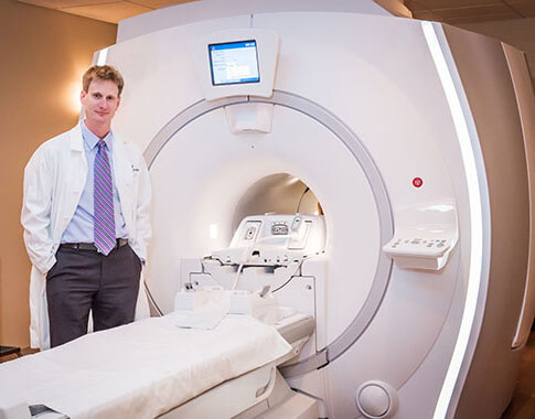
PET/MRI is a hybrid imaging technology that is serving critical diagnostic and treatment purposes for current UCSF patients as well as playing an important role in research for future patients. The PET/MRI technology incorporates the soft tissue morphological imaging of magnetic resonance imaging, or MRI, with the functional imaging of positron emission tomography, or PET, providing high-quality images while reducing patient exposure to radiation.

“MRI is probably the most exciting imaging modality because it’s so flexible, and can be used for many different things,” said Dr. Thomas Hope, assistant professor in residence at UCSF’s Departments of Abdominal Imaging and Nuclear Medicine.
“MRI is used for more than viewing the anatomy,” he explained. “It also characterizes tissues in new ways. These new characterizations include what elements are fluids or solid, the directional flow of liquids, in addition to cellular data. One can also detect different MRI contrast agents in a single scan. Combining these parameters enables an interpretation of the PET data that would not have been possible with previous imaging technologies.”
What is the impact of these advances on patient care? Today, UCSF is the first institution in the United States to diagnose and treat neuroendocrine cancers using PET/MRI imaging based on prostate-specific membrane antigens (PSMA) and the imaging agent edotreotide (DOTATOC).
“We’re honing in on prostate cancer and newer agents that enhance our study of the brain, where you can actually bring in information to combine with the MRI parameters in a way that we could not previously consider.”
Dr. Hope said issues in the pelvis are especially well-imaged with PET/MRI. “These include cervical cancer, the liver, and, we hope in the future, prostate cancer. We’re doing a lot of research projects looking at endocrine tumors and also primary malignancies such as various types of carcinoma and seizure disorders, as PET/MRI is very good for finding the source of seizures. We can inform surgeons and neurologists on how to treat patients with tumors of the brain or squamous cell carcinoma.”
Research on new opportunities combined with continued learning from current uses promises future developments in PET/MRI that will improve patient care. According to Dr. Hope, in the future it will be important to continue to develop new agents for radiotracing, a process in which minute amounts of radioactive material are safely used to enhance scans. They help pinpoint the location of disease, and track metabolic and immune system mechanisms.
“Novel radiotracers will help inform the PET interpretation using these MRI parameters,” said Dr. Hope. “One of the strongest components of MRI is an imaging sequence called diffusion-wave imaging, which allows you to observe cellular lesions. This holds great promise for patients with cancer.”
Thomas Hope, MD, is an assistant professor in residence in the Abdominal Imaging and Nuclear Medicine sections at UCSF and the San Francisco Veterans Affairs Medical Center. In 2007, he received his medical degree from Stanford University School of Medicine and he completed a one-year internship at Kaiser Permanente, San Francisco. From 2008-2012, Dr. Hope completed a residency in Diagnostic Radiology at the University of California, San Francisco, followed by a clinical fellowship in Body MRI and Nuclear Medicine from Stanford Medical Center in 2013.
