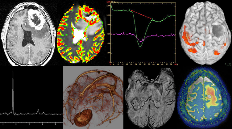
Brain Tumor Imaging at UCSF: On the Cutting- Edge
The following article was written by Soonmee Cha, M.D., Professor of Radiology and Neurological Surgery at UCSF and Principle Investigator, Brain Tumor Research Center.
The brain is the most complex organ in human body. Various tumors arising from the brain are equally complex and comprised of many different types highly variable in biology, response to therapy, and survival outcome. Because the brain is encased in a rigid bony frame of skull, any brain tumor, benign or malignant, is inherently serious and potentially invasive. In addition, there are many brain lesions that are not tumors but can mimic brain tumors and hence require careful differentiation using diagnostic tests for proper medical care. Prompt and accurate imaging diagnosis of the brain tumor is critical for the proper management and counseling of patients with brain tumors and for the selection of specific therapies.
With the help of neurosurgeons, neuro-oncologists, radiation oncologists, neuro-pathologists and imaging scientists, neuroradiologists at UCSF have pioneered the use of Magnetic Resonance Imaging (MRI) to better evaluate not only physical appearance, but functional and biological characteristics of a variety of brain tumors. Using state-of-the art MRI techniques, we are able to capture both high resolution static medical images of brain tumors as well as quantitative functional and chemical changes within brain tumors before, during and after therapy.
With diffusion-weighted MRI, important white matter tracts of the brain can be mapped out in relation to brain tumors so that neurosurgeons can avoid injuring eloquent functioning area of the brain while carefully removing the tumor. Perfusion-weighted MRI provides noninvasive assessment of tumor vascularity that can help differentiate high-grade and low-grade tumors or monitor tumor response to therapy specifically targeting tumor blood vessels. Spectroscopic MRI depicts metabolic derangement within a brain tumor such as the rate of membrane turnover or the status of tumor oxygenation. Relaxometry MRI methods are being investigated to measure the degree of water or edema caused by brain tumor in order to better treat tumor-related complications.
Brain tumor imaging at UCSF combines cutting-edge, high quality anatomic images and quantitative biologic images to impact the clinical outcome of patients with brain tumors by guiding surgical resection, diagnosing tumor-related complications, and monitoring tumor activity during and after therapy.
For more information on neuroimaging with MRI, please see here.


