Measuring Bone Gain and Loss without Biopsy: “Time-lapse” High Resolution Peripheral Quantitative Computed Tomography (TL HR-pQCT)
 Galateia Kazakia, PhD, and researchers in the Bone Quality Research Lab at UCSF are validating a ‘virtual bone biopsy’ using time-lapse High Resolution peripheral Quantitative Computed Tomography (HR-pQCT) for its potential to replace surgical bone biopsy. Currently, surgical bone biopsies are the clinical standard to assess bone formation and resorption, and monitor disease activity and treatment response, but they are invasive, painful, expensive, and cannot monitor disease over time. Furthermore, surgical bone biopsies provide data from a single point in time and so cannot map the varying topology of gain and loss across different locations on the bone which limits understanding of patient response to treatment. Once validated, time-lapse HR-pQCT virtual bone biopsy will be applicable to any study of bone metabolism, enabling precise determination of location and rate of bone gain and loss, which is currently impossible to detect in living humans.
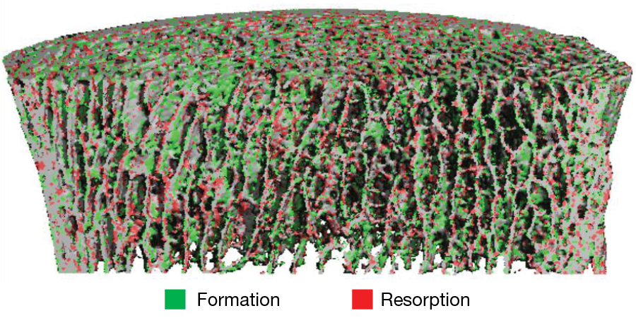
HR-pQCT is a non-invasive, low radiation dose imaging modality that quantifies bone morphologic features with resolution down to 30 µm. Kazakia and her colleagues have explored a novel measure of bone turnover by applying “time-lapse” HR-pQCT in which bone is imaged twice over a longitudinal time-lapse period. Spatial alignment and superposition of the two images enables identification of precise areas (Fig. 1) of bone gain (formation) and loss (resorption) as well as a complete assessment of turnover type (high vs low turnover).
By comparing two time-lapse images produced by the newest generation of high-resolution scanners, this imaging can produce a ‘virtual bone biopsy’ and replace the traditional surgical option. It could allow an unprecedented precise monitoring of the location and rate of bone gain and loss which is currently impossible to detect in patients. When clinicians are able to watch bone gain and loss across load-bearing and non-load-bearing sites, effectively in real time and throughout treatment, it will represent a revolution in patient care for those who suffer from bone diseases such as osteoporosis.
To learn more about this and other imaging research that addresses bone health and pathology, visit Dr. Kazakia’s Bone Quality Research Lab and the interdisciplinary Musculoskeletal Quantitative Imaging Research (MQIR) group.