Using Radiology to Diagnose GERD & Gastric Cancer
The following article was written by Benjamin M. Yeh, M.D., Professor in Abdominal Imaging at UCSF & Assistant Chief of Radiology at the San Francisco Veterans Affairs Medical Center.
The month of November is host to National Stomach Cancer Awareness Month and Gastroesophageal Reflux Disease Week. Although the incidence of gastric cancer has declined over time, it still remains the second leading cause of cancer deaths worldwide. Gastroesophgeal reflux disease, or GERD, is a chronic disease of the stomach and esophagus, affecting up to 1 in 5 or more U.S. adults. Radiology plays an important role in diagnosing gastric cancer and GERD.

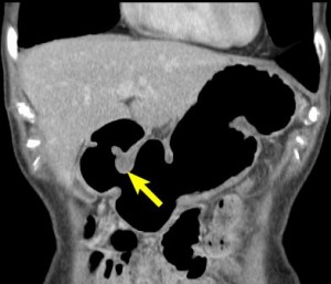
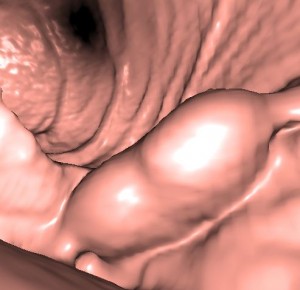
In recognition of awareness for these diseases, I want to highlight several specialized radiological methods offered at UCSF. These methods are geared toward non-invasive imaging of the stomach and esophagus. These imaging procedures utilize oral effervescent granules, which naturally form a small amount of carbon dioxide gas in the stomach to provide gaseous distension of the stomach as a means to obtain a detailed look at the stomach wall at CT and fluoroscopy. From CT image sets, three-dimensional reformations are generated to identify or stage tumors. These reformations allow us to take a more detailed look beyond the bowel mucosa to search for inflammation or lymph node enlargements in nearby areas of the abdomen.
At UCSF, physicians from a number of different specialties work together to diagnose and treat abdominal disease. Radiologists who specialize in Abdominal Imaging collaborate with gastroesophageal surgeons, gastroenterologists, and speech pathologists to evaluate a range of functional and mechanical causes of swallowing difficulties including reflux disease and other sources of abdominal pain.
Click here to learn more about the diagnostic imaging services, including the UGI Series & Barium Swallow, offered at UCSF.

