Multi-Parametric Prostate Cancer Staging Exam

UCSF is the world leader in prostate cancer imaging, based on its extensive NIH-funded research program. The program focuses on the development and clinical translation of advanced prostate cancer imaging techniques, including mult-parametric MRI prostate cancer imaging. UCSF has been instrumental in the development and application of this technique for clinical uses. In fact, this advanced FDA-approved imaging exam has been performed in over 7,000 patients at UCSF, and our radiologists have more than 30 years of expertise in interpreting the data. That translates to the proficiency necessary for best informing the patient of treatment options.  

Multi-parametric MRI prostate cancer imaging exams are performed after a biopsy-proven diagnosis of prostate cancer to provide patients with more detailed information about their disease so they can make the most informed treatment decision and/or understand whether the treatment they have received thus far has been effective.

The UCSF standard multi-parametric MRI prostate cancer staging exam includes:
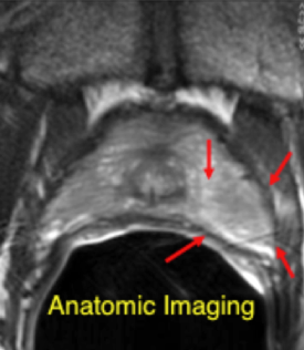
Anatomic Imaging

Prostate cancer can be identified on anatomic imaging (T2 - weighted imaging) based on pathologic changes within the prostate. Anatomic imaging is an excellent technique for assessing the spread of cancer outside the gland. While anatomic imaging can identify cancer within the gland with high sensitivity, the image contrast is not very specific. As a result, it is important to obtain other functional imaging measurements to most accurately identify cancer within the prostate.
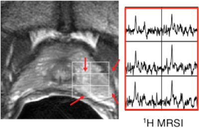
anatomic imaging

Metabolic Imaging

Metabolic imaging, also called MR spectroscopy imaging or 1H MRSI, measures concentrations of important chemicals (metabolites) within the body. It has been shown to improve the identification and characterization (location, size, and aggressiveness) of prostate cancer in patients.

metabolic imaging

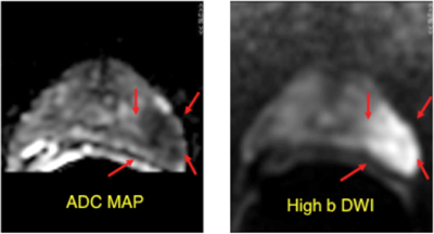
Diffusion Weighted Imaging (DWI)

DIffusion Weighted Imaging (DWI) can be used to assess prostate cancer presence, spatial extent and aggressiveness. The assessment of prostate cancer is possible because DWI is sensitive to the motion of water molecules in tissue at microscopic spatial resolution.

diffustion weighted imaging

Dynamic Contrast Enhanced (DCE) MRI

Dynamic Contrast Enhanced (DCE) MRI is performed by injecting a FDA-approved Gadolinium-based MR contrast agent into the patient and measuring the changes in uptake and washout of the contrast agent, which is increased in prostate cancer. DCE can also be used to assess prostate cancer presence, spatial extent and aggressiveness.

Dynamic Contrast Enhanced MRI

What you should know about a Multi-Parametric Prostate Cancer Staging Exam 

Before Your Exam

In advance of your scheduled exam, please review the following information, including contraindications, safety concerns, and possible contrast agent (if used) nephrotoxic risks (pdf). If ordered by your primary care physician, you may be asked to have a creatinine test done anytime from six weeks up until the same day of the exam.

On the day of the exam, a staff person will be available to answer any questions. You will also be asked to sign a consent form (which is available here to be reviewed at your convenience prior to your appointment - pdf). This will enable us to use the most advanced technology only available at UCSF.

During the procedure, you will be asked to lie down on the MRI platform and turn on your side with your back to the nurse. The nurse will complete a brief digital exam to assess the area for safe probe insertion and then insert an endorectal probe, which is lubricated with KY gel.  Then, you will need to turn on your back. You will be given earphones or earplugs to help block out noise from the MRI scanner. Then, you will be positioned within the bore of the MRI scanner.

While inside the MRI bore and during the course of the scan, you, doctors, and technologists may communicate via a microphone and room loudspeaker system.  In order for us to obtain the best images and data, you will need to remain motionless and relaxed during the entire exam, which lasts about 60 minutes. Note: Ending the exam too soon or having too much body movement during its course can affect data acquisition and render imaging results uninterpretable to the radiologist. 

Upon completion of the exam, you will be moved outside of the MRI bore and the endorectal probe will be removed. After this is done, you may return to the changing area, retrieve your personal belongings, and leave the imaging center. If you should have questions, concerns or experience a non-emergency problem during the exam, you will need to wait for a break between image acquisition sequences to address these to the technologist in the control room.

Note: You will not be exposed to harmful radiation during these studies, nor will invasive procedures, such as a biopsy, be performed.

If you have any questions before your exam, please call (415) 353-4030.

Scheduling

To schedule or reschedule an appointment, please call Radiology Central Scheduling at (415) 353-3900.

Billing and Insurance

Patients, physician office administrators, or case managers should address billing and insurance questions to (415) 514-8888.  Additional information is available on our Billing and Insurance page

Locations

We provide prostate cancer imaging at three locations:

If your exam is scheduled for the Mission Bay Campus, then you must first check in at the China Basin Campus. Personnel are available to escort you from this site to the Mission Bay Campus where the exam will take place.

Exam Results

When the exam has been completed and the radiologists have interpreted the results, a final report will be sent to your referring physician (normally within 3 working days). You may also request a copy of this report, as well as images (provided on CD). Please call (415) 353-9498.

Questions or Concerns?

If you have any questions before your exam, please call (415) 353-4030.