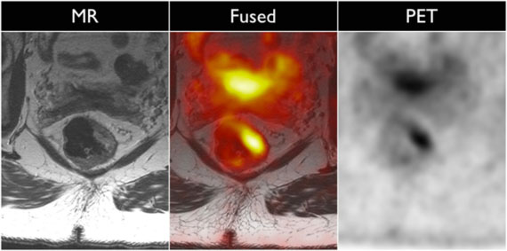
Rectal Cancer PET/MRI
Oncologic staging for patients diagnosed with rectal cancer involves imaging of the primary tumor using MRI as well as evaluation for metastatic disease. This can be performed either using a CT of the chest/abdomen/pelvis or a whole body PET/CT. With the introduction of a simultaneous PET/MRI system, evaluation of the primary tumor using MRI and whole body staging for metastatic disease can be performed on the same machine. This allows for improved workflow and patient convenience. Improved therapy response prediction using information from both PET and MRI data is currently being evaluated, but holds promise for the future.

