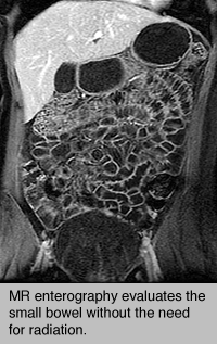
 An MR enterography procedure uses magnetic resonance imaging (MRI) technology to obtain detailed images of the small bowel. MR enterography, also called Magnetic resonance enterography (MRE), is a complementary advanced, accurate and noninvasive diagnostic imaging test to evaluate a broad range of disorders including Crohn's Disease.
The procedure is painless, and there are no known risks, provided the patient has no metal in or on their body and is not pregnant.
In addition to regular preparation for an MRI exam, prior to MR enterography the patient is given two bottles of a special liquid to drink (one bottle 20 minutes before the exam and one bottle 10 minutes before the exam). The liquid serves to distend the bowel and marks the bowel for clear identification during the imaging study. Towards the end of the exam the patient is given a small dose of glucagon followed by an injection of gadolinium (an MRI contrast agent). Glucagon prevents the bowel from moving for a short time, which improves the quality of the images.