Vascular Access and use of Central Lines and Ports in Pediatrics
Intravenous Administration of Contrast Agents for Enhanced CT or MR Scans in Pediatrics
- Peripheral IVs
- PICCS (peripherally inserted central catheters)
- Chest Ports
- Central Lines
- Hand injection into a central line
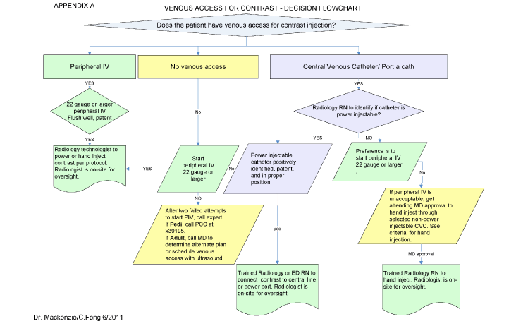
Safe intravenous access, for the injection of intravenous contrast, is vital in obtaining high quality contrast enhanced or angiographic studies. Proper technique is used to avoid the potentially serious complications of contrast media extravasation, air embolism, and damage to the catheter. When the proper technique is used, contrast medium can safely be administered intravenously by power injector, at high-flow rates of up to 2 mls/second (depending on size of patient). A short peripheral IV catheter in the antecubital or forearm area is the preferred route for intravenous contrast administration. However other routes may need to be used and each is considered separately below. The follow flowchart will assist in the decision of obtaining proper venous access for contrast administration.
 Determine contrast administration technique by vascular access type.
(Click on image to view larger)
1) Peripheral IVs
- Pediatric inpatients should be sent to the Radiology Department with a peripheral IV (PIV) or a power-injectable central venous catheter (CVC) that is suitable for the rapid infusion necessary to inject the contrast for the exam. See Nursing policy “Preparation for a Radiologic study with IV contrast (pediatric/neonatal)”.
- The RN responsible for the patient will verify adequate IV access prior to transporting the patient to Radiology. The RN will communicate with the Radiology technologist any pertinent information, including the patient’s IV access. The PIV must be able to tolerate a 1-3 mL per second rapid bolus of NS prior to sending patient to Radiology. If the patient has pain with a rapid infusion of NS, the PIV is not adequate for intravenous contrast injection. Proper enhanced imaging studies require that contrast material be administered RAPIDLY and with force/high pressure, so it is vital that the proper access be verified prior to sending the patient to radiology.
- If anesthesia will be used for the exam, it is not necessary to start a new temporary PIV prior to anesthesia. This can be done once the child is anesthetized.
- The preferable insertion location is in a large vein, such as the anticubital area, cannulating with a large bore catheter (22 gauge or larger). A 24 gauge catheter may be acceptable in neonates and infants, as long as the criteria for rapid bolus infusion are met. Note: A PIV that is adequate for infusion of IV fluids may not be adequate for a rapid bolus of contrast material, since contrast material is more viscous and flows more slowly through catheters.
- It is not necessary to remove a PIV that is adequate for other uses, but it is necessary to have at least one PIV in place that is adequate for intravenous contrast administration before sending the patient down to Radiology for the study. If there are multiple IVs, indicate/mark the PIV that has been verified as adequate.
2) PICCS (peripherally inserted central catheters)
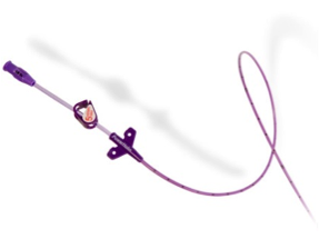
PICCs that are power injectable are clearly marked "power injectable" and have a maximum flow rate printed on the catheter lumen or hub itself. They can be power injected by a trained MD or RN and should only be used according to manufacturer's guidelines in the presence of appropriately trained personnel. These lines include, but are not limited to the following:
Power PICC SV by BARD is a 20 gauge 3 French PICC line that is power injectable up to 1 ml/sec.

A power injectable catheter can be used to administer contrast intravenously.
The Radiology RN will verify markings on the PICC line that identifies it as power injectable, ie. “max mL/sec” on the lumen or “Power Injectable”. Review of the patient medical record may be helpful to determine if the catheter is power injectable. If the catheter cannot be identified as power injectable, it may not be used for power injection.
The Radiology RN/MD will also verify the tip location. The tip must be located in the superior vena cava or at the superior cavoatrial junction. Unacceptable locations include tip pointed wrong way in vein (retrograde the flow of blood) or located in a small or accessory vein. A repeat chest radiograph must be obtained to verify location of the tip (the tips can move) if the current documentation is >1 month old.
3) Chest Ports
- The Smart Port by AngioDynamics is a subcutaneous indwelling central venous access port that is FDA-approved for power injection of intravenous contrast. It has distinctive scalloped edges that can be palpated or seen on a CXR or scout view. Note the “CT” is visible on x-ray image of the newer models of ports as an identifier that this port is power injectable. It is indicated for power injection of contrast media up to 5 mL/sec. and 300 psi pressure limit setting, when used with a Gripper Plus Huber needle. They are MRI conditional at 3 Tesla. This is the most common adult chest port currently placed at UCSF.

Contrast can be administered via chest ports.
- Smith Medical has power P.A.C. port that is power injectable up to 5 ml/sec. when used with a Gripper Plus power P.A.C. Huber needle. They are MRI conditional at 3 Tesla. There are single-lumen and dual-lumen ports that are power injectable. Note that the word “CT” is visible on a x-ray image of the newer models of ports as an identifier that this port is power injectable.
4) Central Lines
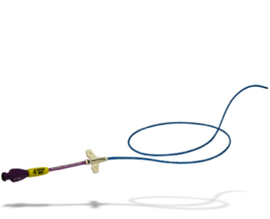
Power Hohn by BARD is a 5 French single lumen catheter that is power injectable.

An intravenous injection of contrast can be given through central lines.
A trained Radiology RN/MD will help the imaging technologist connect the power injectable CVC to the power injector when the following conditions are met.
- The 5 rights for medication administration are satisfied. Check patient identification to ensure the Right Patient, check the Right Contrast (medication), check the Right Dose, check the Right Route, and check the Right Time.
- The tip of the catheter is located at the SVC or at cavoatrial junction.
- Visual inspection of the external portion of the catheter for integrity and proper position.
- Catheter flushes properly and withdraws properly.
- The technologist will perform the power injection after the power injector has been connected to the power injector catheter by the radiology nurse/MD.
The maximum flow rate and psi for Pediatric is 2 mL/sec at <300 psi. The injection rate will be dependant on the specific catheter type, size, and specific exam protocol.
5) Criteria to HAND INJECT through a non-power injectable CVC:
Please see the flow chart at the beginning of this document to help in the decision making for IV access for radiology studies. Placement of a new peripheral IV is the preferred choice of access in patients with non-power injectable CVCs. For pediatric patients without peripheral IV access, a discussion between the patient’s primary attending and the radiologist must also take place regarding the lack of alternate access. A provider order must be written to utilize a non-power injectable CVC for intravenous contrast administration.
1) The Radiology nurse will determine the catheter type and size by visual inspection and review of documentation.
Pediatric patients with non-power injectable central lines, in whom peripheral IV access cannot be obtained, may undergo contrast injection for a CT/MRI by hand injection into the following catheters:
- BARD tunneled 4.2 SL, 6.6 SL
- BARD tunneled 7.0 DL, 9.0 DL (RED port will be utilized)
- COOK non-tunneled 4.0 DL, 5.9 DL, 7.0 DL (distal port will be utilized)
- BD 20 gauge (3 F) PICC non power injectable.
- Other catheters may be acceptable but only after discussion with the radiologist responsible for the interpretation of the study.
2) Hand injection of contrast is an acceptable form of administration if the examination does not require arterial phase of injection (CTA).
3) A trained Radiology RN/MD will perform the hand injection into the non-power injectable CVC when the following conditions are met.
- The 5 rights for medication administration are satisfied. Check patient identification to ensure the Right Patient, check the Right Contrast (medication), check the Right Dose, check the Right Route, and check the Right Time.
- The tip of the catheter is located at the SVC or at cavoatrial junction.
- Visual inspection of the external portion of the catheter for integrity and proper position.
- Catheter flushes properly and withdraws properly.
4) The trained Radiology RN will hand inject contrast per protocol/order followed by 10 mL Normal Saline (0.9 sodium chloride) flush and perform catheter maintenance per UCSF guidelines.
