Joint PICT & ci2 mini Symposium
Date
Type
Time Duration
Speakers

Director, Body RIG
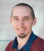
Peder Larson, PhD, is an Associate Professor in Residence and a Principal Investigator in the Department of Radiology and Biomedical Imaging at the University of California, San Francisco. Dr. Larson's research program includes a range of imaging technology development targeting improved clinical outcomes, with projects on metabolic MRI, lung MRI, myelin imaging, PET/MRI, and radiation therapy planning:
Metabolic imaging methods using hyperpolarized carbon-13 MRI
This technology uses non-toxic, non-ionizing contrast agents to provide unique metabolism information, and is currently in clinical trials. Our team develops data acquisition, image reconstruction, and data analysis methods for this technology for a broad range of collaborators and applications. We are also leading new patient studies of this technology in renal cancers and heart disease.
Lung MRI
Conventional MRI performs very poorly in the lungs but our team has developed methods based on ultrashort echo time (UTE) MRI to provide high quality, high-resolution lung MRI. We are pursuing development of functional imaging biomarkers and translation onto clinical MRI scanners, with a focus on pediatric studies to reduce radiation dose compared to CT.
Myelin MRI
Our team has pioneered a new MRI approach for direct imaging of myelin based on ultrashort echo time (UTE) MRI. Other MRI methods provide indirect myelin measurements based measurements on nearby water. We are continuing to develop this technique as a quantitative imaging method, and translating into studies evaluating myelination in disease.
PET/MRI
Hybrid PET/MRI systems combine the functional information from PET tracers with the soft-tissue contrast from MRI. Our team is working on PET/MRI technology developments for motion management, quantitative imaging, and multi-modal data analysis with this modality.
Radiation Therapy PLanning
Applications of MRI in radiotherapy have increased significantly over the past decade due to the high level of soft tissue provided, often allowing for better visualization of tumors and organs at risk versus computed tomography (CT). Our team is working to develop specialized MR techniques to accurately estimate parameters used for radiotherapy dose calculation as a critical step towards MRI-only treatment planning.
Dr. Larson is an active member of International Society for Magnetic Resonance in Medicine, the Institute for Electrical and Electronics Engineering, the UC Berkeley and UCSF Graduate Group in Bioengineering, the California Institute for Quantitative Biosciences, and the Bakar Institute for Computational Health Sciences.

Our research laboratory creates multi-omics prediction models for personalized cancer diagnosis and treatment. I serve as the principal investigator for the MEDomics Consortium (www.medomics.ai). Our group has active research activities in quantitative imaging, natural language processing and the creation of prognostic machine learning models. Our informatics efforts use a wealth of digital information to build point-of-care artificial intelligence tools aimed at assisting physicians in the clinic. Novel algorithms are being developed to produce interpretable multi-criteria analyses tools.

Modality Director, MRI
Javier Villanueva-Meyer, MD, is an Assistant Professor of Clinical Radiology in the Neuroradiology subspecialty section in the Department of Radiology and Biomedical Imaging at the University of California, San Francisco. He received his medical degree from Baylor College of Medicine in Houston, Texas, and he completed his one-year internship at Virginia Tech Carilion School of Medicine in Roanoke. Dr. Villanueva-Meyer completed his four-year diagnostic radiology residency at the UCSF, followed by a Neuroradiology fellowship at UCSF. He also completed a NIH T32 post-doctoral fellowship in the UCSF Department of Radiology and Biomedical Imaging.
Dr. Villanueva-Meyer’s interests focus on advanced MR imaging and imaging based tools to better delineate and characterize brain tumors. His research also involves molecular imaging where he works with collaborators within and outside of our department to develop and implement the use of PET radiotracers in both brain tumors and spinal infection.
Dr. Villanueva-Meyer has experience in CT, MRI and plain film radiography of the brain, head and neck, spine, and peripheral nervous system in both the inpatient and outpatient settings. As well as diagnostic and therapeutic neuroradiology procedures, including percutaneous biopsy, lumbar puncture, myelography, cisternography, epidural and selective nerve root injection.

BIOGRAPHY
Valentina Pedoia, PhD, is an Assistant Professor in the Radiology and Biomedical Imaging Department. She is an Imaging scientist with a primary interest in developing algorithms for advanced computer vision and machine learning to improve the usage of non-invasive imaging as diagnostic and prognostic tool. She obtained her doctoral degree in Computer Science working on features extraction from functional and structural brain MRI in subjects with glial tumors. After graduation, in 2013, she joined the Musculoskeletal and Imaging Research Group at UCSF as post-doctoral fellow to study degenerative joint diseases with compositional MRI techniques.
Dr Pedoia joined UCSF as Faculty In 2018. She is part of the Center of Intelligent Imaging where she serves as Co-director of the Educational Pillar. Her current research is focused in exploring the role of machine learning to extract imaging biomarkers of several musculoskeletal conditions including knee and hip Osteoarthritis, shoulder Instability and lower back pain. She develops analytics to model the complex interactions between morphological, biochemical and biomechanics aspects of the joints as a whole. Her goal is to develop efficient and effective data-driven models that able to extract imaging features and use them to identify risk factors, stratify patients and predict outcomes.
She has great interest in the clinical translation of novel technology, as such she is invested in making the image acquisition and processing faster, safer, and smoother for patients and clinicians.

Radiology
Scientific software developer with a focus on data visualization, algorithms and image processing. An advocate of open-source software with a focus on building tools to apply new technologies to cutting-edge science.
