Breast Cancer Awareness Month Research Roundup

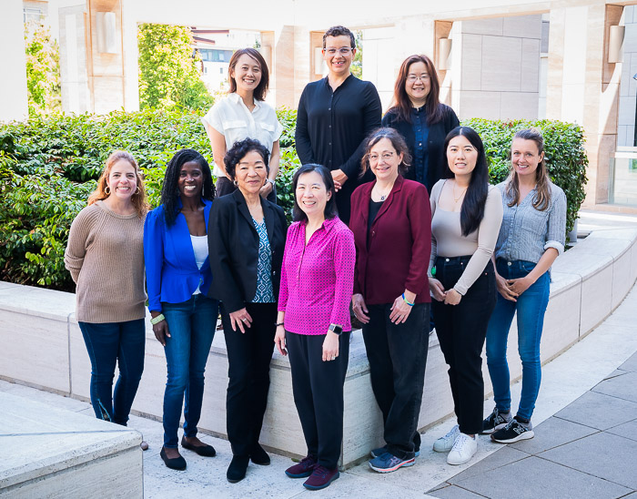
October is Breast Cancer Awareness Month and an opportunity to recognize the researchers and clinicians who enable care for patients suffering from this disease.
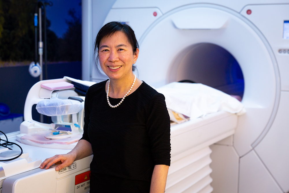
UCSF Radiology and Biomedical Imaging scientists and doctors are improving how we diagnose, monitor, and treat breast cancer with AI, advanced imaging, and enhancements to the patient experience.
Recent Work from Our Colleagues
An Algorithm to Predict Treatment Success and Enable Early Surgery
A phase 2 clinical trial published in Nature Medicine designed a way to rapidly assess the effectiveness of novel drugs for patients with high-risk, early-stage breast cancer and allow them the option of early surgical resection if they are likely to have achieved a pathologic complete response.
Over 12 years, the trial evaluated 23 drugs and drug combinations, with some, such as pembrolizumab, going on to phase 3 trials. By assessing over 2,100 patients with serial MRIs during neoadjuvant therapy obtained in these initial studies, the investigators developed a model to predict pCR that, in combination with core biopsy, could potentially identify a group of patients that could safely go to surgery before completing an entire planned neoadjuvant regimen, thereby safely avoiding further toxicity. This combined algorithm is used to make recommendations in the ongoing I-SPY 2.2.
Wen Li, PhD, Natsuko Onishi Yamashita, MD, PhD, Elissa Price, MD, and Nola Hylton, PhD, of the Breast Imaging Research Group, with senior author Laura Esserman, MD, MBA, professor of surgery in the School of Medicine collaborated with colleagues at 27 other universities and healthcare institutions.

AI Breast Cancer Screenings Require Diverse Training Sets
A research highlight published in Radiology, reviewed the retrospective cohort study by Derek Nguyen and Yinhao Ren et al. from Duke Medicine appearing in Radiology.
The study revealed significantly higher false-positive case scores in Black patients and lower in Asian patients compared with White patients. False-positive risk scores were higher in Black patients, patients aged 61 to 70, and those with extremely dense breasts, compared to White patients, patients aged 51 to 60, and patients with fatty breasts. These findings underscore the need for greater transparency of algorithm development processes and the necessity of using diverse data sets for algorithm training.
First author Nour Homsi is an alumna of the RIDR program and was advised by senior author Maggie Chung, MD.

Can Synthetic Mammography Replace Digital Mammography?
A recent survey of practicing radiologists, published in the Journal of Breast Imaging, assessed anonymized respondents’ demographics, current mammographic screening protocol, confidence interpreting synthesized mammography (SM) for mammographic findings, and perceived advantages and disadvantages of SM.
SM creates an image formatted like a traditional 2D mammogram with data from a 3D digital breast tomosynthesis (DBT) scan. These synthetic 2D mammograms have greater compatibility with existing infrastructure, match existing regulatory guidelines, and allow for easier comparison to previous mammogram records.
For most survey respondents, SM has replaced Digital Mammography in DBT screening. Most respondents felt confident using DBT/SM to evaluate masses, asymmetries, and distortions. The radiologists routinely interpreting DBT with SM alone reported higher confidence, fewer disadvantages, and more advantages with SM, suggesting overall satisfaction with SM.
UCSF Chief of Breast Imaging Bonnie Joe, MD, PhD, joined colleagues from Cornell, Harvard, University of Texas, and Duke University to analyze these results.

Patients are Satisfied with Contrast-Enhanced Mammography
In a research highlight from Radiology, Sofia Fung-Lee and Bonnie Joe, MD, PhD, commented on a recent study by Matthew Miller et al. in the Journal of Breast Imaging. Fung-Lee and Joe described the acceptance of and appreciation for Contrast-Enhanced Mammography (CEM) among patients undergoing breast cancer screening, reporting minimum unpleasantness, willingness to continue with CEM, and a willingness to recommend CEM screening to a friend. The study showed that screening CEM can be well received among patients with dense breasts, with study participants reporting high satisfaction. This was especially true when CEM could be administered at the same cost as traditional screening mammography.
Dense breasts limit the sensitivity of traditional mammography, an obstacle which CEM can overcome. The study, conducted at an outpatient screening clinic, showed that patients are receptive to this new modality as a routine screening examination.

