Biomagnetic Imaging Laboratory
The Biomagnetic Imaging Laboratory (BIL) is dedicated to advancing our understanding of brain function, particularly speech and language, through non-invasive imaging. We utilize Magnetoencephalographic (MEG) imaging, alongside Functional Connectivity (fMRI), Electrocorticography (ECoG), and Transcranial Magnetic Stimulation (TMS). Beyond groundbreaking research into brain network dynamics, we provide vital clinical services, including pre-operative brain mapping for neurosurgeons to protect eloquent function in patients with tumors and the localization of epileptic zones in patients with epilepsy. We’re one of only two clinical MEG sites in California, and among just 25 sites in the United States.
For Referrals – How to Refer a Pateint
For Patients
Research Areas
Development and Validation of Algorithms and Tools for Electromagnetic Source Imaging
 Magnetoencephalographic Imaging (MEGI)
The principal goal here is to improve the use of electromagnetic source imaging (ESI is the combined use of MEG, EEG and MRI) in clinical and research practice through the development of better algorithms for image reconstruction and analysis. Another goal is the development of tools for integration of ESI with other imaging modalities such as DTI and fMRI. Ongoing projects include the development of various algorithms for selective signal cancellation, separation and localization of brain sources from EEG and MEG data. We draw upon advances in statistical signal processing, machine learning, and probabilistic Bayesian inference and apply such techniques to analyze MEG and EEG data. Another ongoing effort is to develop algorithms for automated detection and localization of epileptogenic zones. All Algorithms are validated by comparing ESI reconstructions with electrocorticography (ECoG) recordings in patients with brain tumors and in patients with epilepsy. Algorithm validation is also performed in animal models by comparing imaging data such as ESI reconstructions and DTI connectivity with electrophysiological and neuroanatomical measurements. Clinical applications of this work will be tested in patients with brain tumors and in patients with epilepsy.
Determining the Biophysical Basis for Novel Anatomical and Functional Brain Imaging Methods
 Electrocorticography (ECoG)
The goal here is to determine the biophysical basis of auditory and somatosensory cortical activation and connectivity as assayed by modern functional brain imaging methods such as ESI and DTI. In ongoing experiments in patients, with brain tumors or intractable epilepsy, and in animals, we record magnetoencephalography (MEG), electroencephalography (EEG), electrocorticography (ECoG), functional magnetic resonance imaging (fMRI), diffusion-tensor imaging (DTI) data. In animals, we can also record local field potentials and action-potentials from microelectrode arrays and measure neuroanatomical tracer uptakes. Linking such data obtained in the same subjects in response to the same stimuli across these recording methods enables comparisons of imaging across multiple scales and modalities. Current projects include studies of representations of somatosensory and auditory stimuli (in monkeys and cats) and of speech, language and memory responses in humans.
Imaging Cortical Spatiotemporal Plasticity Associated with Learning
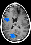
 Resting-state network connectivity predicts recovery in ischemic Stroke.
The goal here is to examine cortical plasticity due to learning and experience in normal adult humans using ESI. We are specifically interested in cortical plasticity in response to dynamic stimuli in the time-scale of tens to hundreds of milliseconds. We have been examining plasticity, associated with perceptual learning, in representations of simple and complex acoustic stimuli as well as speech and language stimuli. We are also examining plasticity of somatosensory and motor representation changes due to training and perceptual learning.
Neuroimaging of Speech, Language and Memory
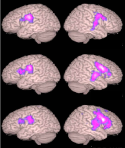
 Resting MEGI Functional Connectivity in Schizophrenia predicts symptoms.
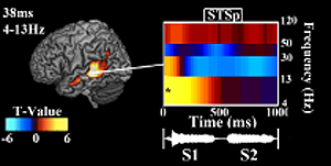
The goal here is to examine the spatiotemporal dynamics of brain networks involved in speech, language and memory processes. We specifically focus on overt speech production, its interaction with auditory feedback processing, and with language and memory processes. Several recent studies have shown that speaking causes "speaking-induced suppression" or SIS - a suppressed response to self-produced speech when compared to identical speech from an external source - in auditory cortex and associative regions. In our own recent work, we have shown that SIS is present in auditory cortex, and does not result from overall inhibition of auditory cortex during speaking. Rather, SIS results from a comparison between actual auditory input and an internal "speaking-induced prediction" (SIP) of that auditory input. What is the functional significance of SIS and SIP? Based on several lines of evidence, we have developed a conceptual working model for SIS and SIP. The principal goal of this research is to test predictions from this model. Our overall approach capitalizes on unique real-time speech feedback alteration methods developed by our research team, the excellent spatial resolution of functional magnetic resonance imaging (fMRI), the excellent temporal resolution of electromagnetic source imaging (ESI) and advanced analyses methods that we have developed for reconstructing spatiotemporal dynamics and connectivity of distributed cortical networks.
Developing Novel Clinical Applications for ESI
 In addition to existing clinical research projects in brain tumor and epilepsy patients, we are also in the process of developing several protocols and procedures for examining novel clinical populations. Ongoing projects including conducting ESI studies on patients with Schizophrenia, Mild Cognitive Impairments, Parkinson's disease, Autism, Traumatic Brain Injury, Stroke, Focal-Hand Dystonia and Agenesis of the Corpus-Collosum.
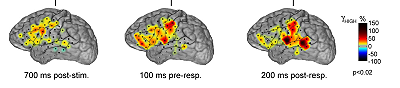
Center versus peripheral vowel productions in a sample subject.
A, Acoustic variation across repeated productions, shown in 2D formant frequency space. Green represents center productions; red represents peripheral productions; black represents remaining productions.
B, A source localization algorithm (Owen et al., 2012) determined the coordinates and field strength of the M100 peak (MNI56,24, 8 in this subject).
C, MEG traces aligned tovowel onset, separated into center and peripheral trials as determined by A. Shaded regions represent SE (n100); vertical bars on the y-axis represent SIS magnitude.
The Biomagnetic Imaging Laboratory (BIL) provides the following:
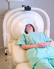
Measure magnetic and electric brain activity
 • Clinical patients for presurgical work up (brain tumor, epilepsy)
• Research patients with various neurologic conditions (traumatic brain injury, dementia, autism, schizophrenia, stroke, tinnitus, spasmodic dysphonia, focal hand dystonia, primary progressive aphasia, agenesis of the corpus callosum, dyslexia, etc.)
MEG/MSI Support and Service for Clinical & Research Purposes
 • MEG - Magnetoencephalography
• MSI – Magnetic Source Imaging (MEG + MRI = MSI)
Perform Source Analysis
 • Standard analysis software/protocols
• Specialized source analysis algorithms developed in house
Administrative Support
 Coordination of patient schedules, scheduling, screening, consenting, recharging, CHR management, reimbursements, etc.
Team Members
Clinical Operations Team
- Srikantan Nagarajan, PhD – Director
- Heidi Kirsch, MD, MS – Clinical Director
- Robert Knowlton, MD – Clinical Director
- Corby Dale, PhD, MPH – Staff Scientist
- Leighton Hinkley, PhD – Staff Scientist
- Velmurugan Jayabal, PhD – Staff Scientist
- Anne Findlay – Lab Manager
- Gavin Belok – MEG Tech
- Joshua Chon – MEG Tech
- Srivatsan Tennathur – MEG Tech
- Natalie Brunwin – Clinical Research Coordinator
- Dylan Davis – Clinical Research Coordinator
- Saloni Gupta – Clinical Research Coordinator
- Rachel Lentner – Principal EEG Tech
Research Team
- Srikantan Nagarajan, PhD – Director
- Corby Dale, PhD, MPH – Staff Scientist
- Leighton Hinkley, PhD – Staff Scientist
- Velmurugan Jayabal, PhD – Staff Scientist
- Anne Findlay – Lab Manager
- Kurtis Brent – Graduate Student
- Alvincé Pongos – Graduate Student
- Zooey Zhang – Graduate Student
- Jessica Gaines, PhD – Postdoc
- Pooja Prabhu – Postdoc
- Rabab Rangwala – Postdoc
- Saloni Gupta – Clinical Research Coordinator
Advisory Committee
- John Houde, PhD – Faculty Collaborator
- Kamalini Ranasinghe, MD, PhD – Faculty Collaborator
- Karuna Subramaniam, PhD – Faculty Collaborator
- Carly Demopoulos, PhD – Faculty Collaborator
- Ashish Raj, PhD – Faculty Collaborator
- Shawn Hervey-Jumper, MD – Faculty Collaborator
- Marilu Gorno-Tempini, MD, PhD – Faculty Collaborator
- Steven Cheung, MD – Faculty Collaborator
- Phiroz Tarapore, MD – Faculty Collaborator
- Heidi Kirsch, MD, MS – Clinical Director
- Corby Dale, PhD, MPH – Staff Scientist
- Leighton Hinkley, PhD – Staff Scientist
- Velmurugan Jayabal, PhD – Staff Scientist
- Anne Findlay – Lab Manager
Alumni
- Joshua Bear, MD – University of Colorado, Denver
- Ethan Brown, MD – UCSF
- Edward Chang, MD – UCSF
- Elizabeth Disbrow, PhD – Louisiana State University, Shreveport
- Dario Englot, MD, PhD – Vanderbilt University
- Adrian Guggisberg, PhD – University of Geneva
- Ilana Hairston, PhD – Tel Hai Academic College
- Kenneth Hild, PhD – Amazon.com
- Kitti Kaiboriboon, MD – Case Western Reserve University
- Tal Kenet, PhD – Harvard Medical School – Massachusetts General Hospital
- Juan Martino, MD, PhD – Hospital Universitario Marques de Valdecilla
- David McGonigle – Cardiff University
- Caroline Niziolek, PhD – Boston University
- Maneesh Sahani, PhD – Gatsby Computational Neuroscience Unit, University College London
- Carsten Stahlhut, PhD – Technical University of Denmark
- Noriko Tanigawa – University of Oxford
- Virginie Van Wassenhove – Neurospin, France
- Maria Ventura, PhD – UCSF
- Kelly Westlake, PhD – University of Maryland, Baltimore
- David Wipf, PhD – Amazon AI Lab in Shanghai
- Zhao Zhu, PhD – South San Francisco
Past Graduate Students
- Sarang Dalal, PhD – Aarhus University
- Alexander Herman, MD, PhD – UCSF
- Naomi Kort, PhD – UCSF
- Julia Owen, PhD – UCSF
- Johanna Zumer, PhD – University of Birmingham