
Right Time, Right Place: Radiology Makes the Difference for Patients in Emergency, Acute Care, and Surgery Settings
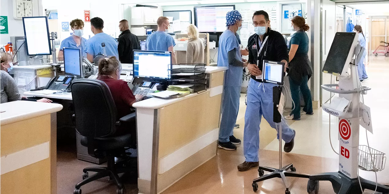
Emergency Radiology and Interventional Radiology bring the service of imaging to the patient, both when and where it is needed. Meeting this need through both capacity and chronology, Mark Wilson, MD, ZSFG Radiologist-in-Chief and Jason Talbott, MD, PhD, Chief of Emergency Radiology are focused on improving patient care by strengthening our ability to handle multiple simultaneous emergencies, and by increasing coverage during late and overnight hours.
The Emergency Radiology division has vastly reduced turnaround times on late night imaging studies, while Interventional Radiology’s collaboration with Trauma surgery, the Neurovascular service, and the emergency department at ZSFG has eliminated dangerous delays between intake and treatment for life-threatening crises. Their evolution represents the Department of Radiology and Biomedical Imaging’s ongoing commitment to patient care and safety.
Emergency Radiology: When They’re Needed

Dr. Talbott leads the Emergency Radiology division at ZSFG and has recently expanded faculty coverage to UCSF’s Parnassus location as they strengthen ties with the pediatric emergency rooms at Benioff Children’s Hospital at Mission Bay and Oakland. He was part of the group who set out to improve UCSF’s ability to supply timely image readings after hours and overnight, while maintaining the autonomy of the call experience for UCSF residents, which is a critical component of their training. With the leadership of Mark Wilson, MD, ZSFG Chief of Radiology, faculty members Jason Talbott, MD, PhD, Shital Gandhi, MBBS, and Amrutha Ramachandran, MBBS, staff this new division along with eight per-diem physicians, and newly hired faculty member Masis Isikbay, MD, who joined the group in September 2024 to cover the Parnassus site. Dr. Christopher Murphy, a recent graduate of the UCSF residency and current abdominal imaging fellow at UCSF, was recently hired and will join the Emergency Radiology division in September of 2025. Talbott is recruiting for one to two additional open positions to cover Parnassus acute care and the emergency department.
Talbott emphasized that resident-led after hours and overnight call shifts are a valuable educational resource, “On-call residents handle the protocol, interpretation and initial wet reads for virtually all of the on-call imaging exams at ZSFG. It’s a great learning environment and something that our resident trainees value greatly. Diagnostic and treatment decisions are made quickly in an emergency department environment, which builds trainees’ expertise, efficiency, and confidence.”
In the absence of Emergency Radiology services, a radiology attending provides a final read for off-hour studies the next morning. With the ever-growing volume and complexity of on-call CT and MRI studies, the need for real-time radiology attending review of these exams has increased. Delayed attending review of ED studies can present a challenge for continuity of care. Talbott says, “Approximately sixteen percent of our patients are undomiciled or unstably housed. So, we often can’t call them back or count on follow up once they have been discharged from the ED.”
UCSF residents do an incredible job on-call and the overall rate of significant call-backs (i.e. cases where the final attending read the next day is discrepant from the on-call resident-provided wet read) is relatively low. Nevertheless, these call-backs can lead to delayed diagnosis and are an issue of health equity that Talbott has worked to solve by developing the Emergency Radiology Division to provide real-time attending reads for the very busy period from 5:00 pm to midnight, rather than requiring patients and providers to wait until the following morning for final reads.
Talbott said, “We had the chance to learn from the challenges of the pandemic. Remote attending work enables flexibility, limits interference with on-call resident autonomy, and allows residents to maintain ownership of call responsibilities. In the future, as we look to expand coverage beyond midnight, it also allows us to leverage time zone differences across the USA to our advantage and more efficiently staff late night and early morning hours.”
The growing ER division provides 100% evening coverage at ZSFG and currently provides coverage with final reads two nights a week for all emergency department cross-sectional imaging studies at Parnassus and Mission Bay campuses. This model has reduced discrepant overreads by more than 55% for patients discharged from the emergency department at ZSFG. Comparing the same periods, the average turnaround time from wet read to final read dropped from nearly 30 hours to approximately 45 minutes. The turnaround time is targeted to be less than 60 minutes.
Interventional Radiology at ZSFG: Where They’re Needed

At a large safety-net hospital and trauma center like Zuckerberg San Francisco General, sometimes everything happens at once. During the summer of 2023, the Interventional Radiology team at ZSFG hospital was faced with the challenge of caring for four patients simultaneously who were experiencing life-threatening stroke, brain trauma, or heart attack, and all in need of immediate care. Fortunately, Mark Wilson, MD, and the radiology and interventional radiology teams at ZSFG had planned to meet such a challenge.
Dr. Wilson took the position as Chief of Radiology and Chief of Interventional Radiology at San Francisco General Hospital in 2008, the year before the city decided to construct a new hospital building for the historic medical campus on Potrero Hill. Work designing the titanic new project began almost immediately, and Wilson was involved in the design process from the beginning.
The mandate for this new hospital was to “bring the service to the patient”, and interventional radiology stood to play a major role. The hospital reflected 150 years of organic growth, as divisions filled the space available based on history rather than a workflow pattern. A patient heading to the operating room and in need of interventional radiology or cardiology might be wheeled off to three different floors.
The new construction represented a chance to position San Francisco General Hospital in the best practices of hospital design and organization. Imaging is now at the forefront, with imaging machines present in the emergency ward, integrated into intake and positioned merely a few direct doors away from the ambulance bays. Wilson also helped with the decision to locate Interventional Radiology within the same space as the operating rooms.
During the design process, Wilson teamed up with James Marks, MD, PhD, Chief of the Medical Staff and Chief of Performance Excellence at ZSFG, to create an environment that integrated the possibilities of interventional radiology with the operating rooms. The primary IR suites are directly across a single hallway from the operating rooms, and one of the primary operating rooms is a hybrid IR room equipped with integrated bi-plane CT machines, retreating to the corners on robotic arms when not needed at the operating table.
The primary IR suite itself is a beautiful arrangement; a symmetrical butterfly of multiple rooms, each half centered around a reading room with broad windows into the arced control room, which looks out into two different procedure rooms. The effect is a reassuring display of technology, like stepping onto the bridge of a starship. Here, patients are easily transferred from biplane imaging to MRI by their bed passing through a single door, crucial when time is of the essence such as in stroke, all observed without break by the technologists and radiologists.
Improving Imaging Workflow for Emergency Radiology and Acute Care Settings

Masis Isikbay, MD, the most recent addition to the Emergency Radiology team, is keen to optimize imaging workflows in the emergency department and urgent care settings.
By further refining clinical guidelines for when it is appropriate to order imaging studies, Isikbay aims to ensure that patients receive the right tests at the right time. The goal of this work is to triage patients appropriately so that those with serious diagnosis receive the appropriate care (with minimal delay).
In urgent/emergency care settings where moments matter, imaging workflow optimization can help providers be confident they are working up the patient appropriately while making sure the correct patients are admitted to the hospital for further evaluation. Isikbay emphasizes, “You cannot do everything all the time because our medical resources are finite. We can, however, leverage clinical data to help refine our approach to make sure we keep our patients safe and minimize the possibility of missed findings.”
Take dizziness, for example. Patients presenting with dizziness or vertigo can have a wide range of underlying causes. These can range from simple dehydration to life-threatening conditions like stroke / vertebral arterial dissection which are dangerous possibilities, but also low probability.
Care providers must strike a balance based on clinical judgement and would benefit from targeted research that can help identify when an imaging study should be ordered and specifically what test would best answer the clinical question. In instances where the imaging workflow is not clearly established, imaging volumes can be elevated unnecessarily, which can contribute to provider/radiologist burnout (without helping drive the patient’s care forward).
Isikbay’s approach involves carefully assessing each patient’s symptoms and medical history to determine the most appropriate imaging tests. He has developed a clinical research workflow which pulls clinical data from APeX and merges it with results from radiology reports in mPower to create a study database. This allows him to more easily study uncommon and dangerous causes – like vertebral artery dissection – for common presentations such as vertigo.
Isikbay uses these databases to identify presentation patterns, diagnostic efficacy of imaging studies and modalities, and glean patient outcomes. With this information, he plans to provide clinical support tools for urgent care/emergency department providers such as clinical screening questionnaires to help identify which patients would most benefit from imaging workup. Additionally, he aims to better establish which imaging study would best workup the patient appropriately.
Isikbay hopes these efforts will help patients feel more comfortable as they understand their care and provide useful guidance for ED physicians. “We can communicate to patients that we know what to look for. If a patient does not have red-flag symptoms, we can better contextualize the urgency of the workup they need. For patients who do have red-flag symptoms, we intend for these support tools to help ED physicians quickly order the correct studies to diagnose and treat truly emergent conditions.”
Vertigo Workup

For the workup of vertigo, Isikbay is developing clinical screening questions with the help of urgent care providers, emergency physicians, and neurologists to help identify those patients who should be imaged for vertebral artery dissection (with a CT angiogram of the head/neck) and those who may have a stroke (a focused stroke protocol MRI of the brain without contrast). While preliminary and pending validation in both internal and external databases, examples of symptoms that likely indicate further workup include:
- Acute rapid onset of vertigo
- Persistent vertigo that does not come and go
- Presence of double vision which is persistent
- Difficulty swallowing or difficulty speaking
- Additional focal neurological deficits (such as new acute limb weakness)
Patients who have some or all of the above symptoms would more likely benefit from additional imaging, while patients with more benign causes of vertigo should, generally, not present with these findings.