
Meet the Postdocs Turning Images into Answers: Part 2
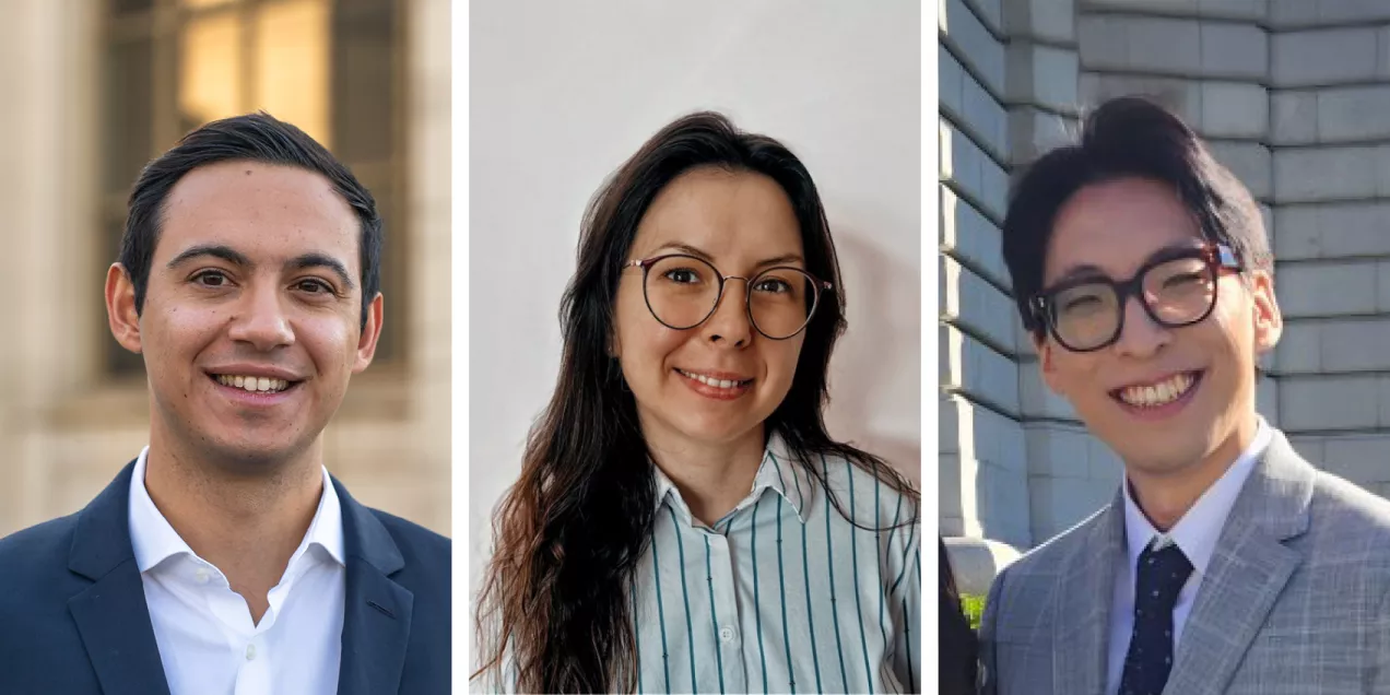
The three postdocs profiled here work on device development for navigating the brain’s venous system, brain metabolism and Alzheimer’s disease, and creating radiotracers to detect bacterial infection. We are grateful for and inspired by their contributions to clinical translation. Don’t miss the postdoc profiles in Part 1.
Panos Pardalidis, MD: Neuroendovascular Surgery and the Venous Side of the Brain

Panos Pardalidis, a specialist in neuroendovascular surgery, studies the brain’s venous drainage. He explains, “the arteries supply the blood the brain needs, but there is also the venous side, which takes that blood back to the heart, and we still don’t know as much about it.” Panos hopes to improve diagnosis and create new treatments for conditions related to this area, among them pulsatile tinnitus, in which a patient hears a rhythmic sound with no source.
His path brought together two interests he was unwilling to choose between. After earning his medical degree in the UK and practicing in London, Panos pursued a master's degree in biomedical engineering at UC Berkeley. “This research at UCSF lets me bring those two interests together, combining medicine, imaging, engineering, and innovation,” he shares. He is most proud of a new diagnostic device his lab is developing, a catheter-like tool that navigates the blood vessels of the brain. After building a working prototype, Panos tested it in a benchtop model that simulates the body’s vasculature and saw promising results earlier this year.
Panos’s advice comes from experience. “It took me a while to realize this is not like most jobs, where if you keep trying, you eventually solve a problem the same day,” he says. He suggests taking a step back when an approach stalls. “Research can involve very complex, unique problems, and sometimes a fresh perspective or discussing the problem with colleagues and your supervisor can make a big difference.”
Tamara Vasilkovska, MD, PhD: Brain Metabolism and Alzheimer’s Disease

Tamara Vasilkovska studies brain metabolism and how it changes with age and in Alzheimer's disease. She explains her move from clinical medicine into research this way: “I wanted to better understand systemic and dynamic changes in the brain in a noninvasive manner, with the idea to explore novel biomarkers, diagnostic tools, or treatments that can help people on a bigger scale.” Working with preclinical models, Tamara looks for the earliest changes in metabolism which may signal an earlier chance to intervene.
She is most proud of a recent finding, made with a technique she describes with enthusiasm. “I was able to show that if you switch a gene that is important for Alzheimer’s disease, you can see a rescue in metabolism, and you can monitor that with hyperpolarized MRI.” Watching that reversal unfold within the lifetime of a single model was, as she puts it, “the best of both worlds.”
Tamara is grateful for the community around her, colleagues and a supervisor who lends a hand even on short notice. “Science would not be what it is if you don’t have that community,” she shares. Her advice to new postdocs is to keep growing. “See your skills as something that needs to be improved and venture outside your comfort zone. A postdoc is the right time to do that.”
Sanghee Lee, PhD: Molecular Imaging Probes for Detecting Infection

Sanghee Lee designs the molecules that make disease visible on a scan. His work involves creating and evaluating imaging probes and radiotracers. His current project develops probes that detect bacterial infection by distinguishing bacteria from the body’s own cells, an advance that could lead to earlier diagnosis and better treatment. “I have always been drawn to invisible things,” he shares, “and I had a strong desire for my research to benefit people’s health in a meaningful way.”
When asked how he explains his work to others, Sanghee offers a fitting analogy. “Our work is very much like a live broadcast. The radioisotopes are short-lived and disappear over time, so we can’t prepare everything in advance like a pre-recorded show. On the day of imaging, every element had to operate in perfect harmony and on a strict timeline.”
As he completes his postdoc and moves into a professional researcher role, Sanghee offers advice drawn from the challenges inherent in scientific inquiry. “Don’t be afraid of failing. Success is valuable precisely because it comes after failure, and the challenges we face are exactly what will allow us to eventually shine.”