
Meet the Postdocs Turning Images into Answers: Part 1
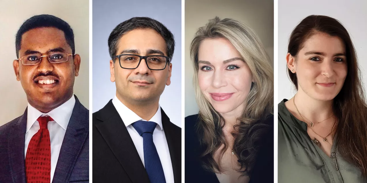
The four postdocs profiled here work on advanced MRI techniques for prostate and breast cancer, neural biomarkers for Alzheimer’s disease progression, and imaging biomarkers for joint degeneration and pain. We are grateful for and inspired by their contributions to clinical translation. Don’t miss the postdoc profiles in Part 2.
Daniel Gebrezgiabhier, PhD: MR Hardware and Hyperpolarized 13C MR Imaging for Prostate Cancer

For Daniel Gebrezgiabhier, a career in imaging began far from any MRI scanner. “The first time I saw an MRI scanner was at UCSF,” he shares. Trained as a biomedical engineer with a focus on medical device development, he was drawn in quickly. “As soon as I started learning about the physics behind radio-frequency engineering, I was fascinated.” Today, Danny develops MR detector hardware and novel hyperpolarized (HP) carbon-13 MRI methods to better detect and stage aggressive cancers within the prostate as well as local metastases (spread) to the prostatic bed and adjacent lymph nodes. When translated to the clinic, Danny hopes his research will more accurately predict outcomes for individual prostate cancer patients and enable clinicians to select the most appropriate treatment.
Danny is especially proud of MRI hardware he helped redesign, which improved sensitivity and image quality by about 80 percent compared to the previously used device. The first time it was used on a prostate cancer patient, HP carbon-13 imaging revealed two suspicious areas conventional imaging had missed. When both spots were later confirmed as cancer Danny observed that, “my device is being used and making a difference.”
His advice to new postdocs is simple. “Keep your curiosity high, use every resource around you, including your colleagues, and make sure you are doing what you love.” Danny is inspired by his faith, his family, and his friends, including those who, despite their ability, never had the opportunities he has had. “I feel like I’m doing this for them too,” Danny shares.
Pouya Metanat, MD, MPH, MS: Breast MRI Quality Control and Personalized Cancer Care

Pouya Metanat is focused on ensuring that a national breast cancer study can trust its own images. UCSF is one of the imaging centers for this national, multicenter trial, which follows patients through chemotherapy with MRI scans taken across more than thirty sites. Because the study sites use different scanners and software, image quality can vary between sites, which can affect the measurements researchers rely on. Pouya aims to understand how much that variability influences the assessment of a patient’s response to treatment.
Coming from a medical background with a master’s degree in computer and electrical engineering, Pouya was drawn to MRI at UCSF where Nola Hylton, his principal investigator, developed many of the breast imaging biomarkers used today. He values the ability to interact with patients directly, which can be rare in research, and often scans patients himself. “That was one of the reasons I went into medicine,” he said, “because I like seeing how I can be impactful in people’s lives.”
For early career researchers, Pouya offered advice that he still practices himself: “Reach out to other people for mentorship. It’s never too early or too late, and it has opened doors for me I never knew were possible.”
Jennifer Senta, PhD: Neural Biomarkers for Predicting Alzheimer’s Progression

Jennifer Senta came to neuroscience from mathematics and actuarial science, drawn by the chance to put her technical skills toward something with real human impact. She found it in Alzheimer’s disease. Her research uses magnetoencephalography, or MEG, to measure changes in brain activity at the earliest stages of cognitive impairment. Diagnosing Alzheimer’s and estimating its current stage are possible today, but as she explains, “It is still very difficult to predict an individual’s expected disease course from the earliest onset of symptoms.” She hopes these early signals, as measured by MEG, can serve as non-invasive biomarkers with prognostic value, guiding diagnosis and treatment for each patient.
Her technical training now informs everything from computational models of neural activity to the signal processing behind MEG, but the motivation is personal. Contributing to solutions for a disease that “robs people of memory and independence,” she says, “feels like both a privilege and a vocation.” She has found a team to match. “The people I work with at UCSF are among the brightest minds I’ve encountered,” she shares, “but more than that, they are also wonderful people who are supportive and collaborative.”
Her advice for new postdocs is grounded in perspective. “Never compare your own academic path to anyone else’s,” she says. “There will always be other researchers with seemingly more publications, larger grants, or higher profiles.” Instead, she suggests focusing on continual improvement and on “how lucky we are to be able to explore our deepest questions for living.”
Tugce Ulas, MD: Muscle Health and Imaging Biomarkers for Joint Degeneration

Tugce Ulas is interested in a part of the picture radiologists often overlook. “As a radiologist, when we look at an MRI, we usually focus on the joint or the spine itself,” she said. “I’m interested in the role of the muscle.” Her research examines skeletal muscle in osteoarthritis and spine degeneration, pairing quantitative MRI with an AI algorithm to measure the amount and quality of muscle and relate it to disc degeneration and low back pain.
One finding she is especially proud of came from her lab’s first longitudinal study of knee osteoarthritis. “We found that the change in thigh muscle volume over time was associated with both structural degeneration of the knee joint and decline in physical function,” she said. “It tells us that muscle can tell us more about the patient’s risk.” The team now hopes to extend the approach to the spine.
What Tugce values most is working alongside colleagues who each bring a different perspective. “Everyone looks at the same problem from a slightly different perspective,” she adds.