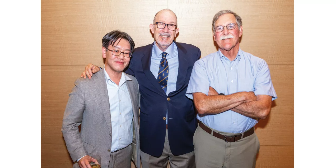
Curious, Devoted, Nature-Lover: William Dillon, MD, Looks Backward and Forward
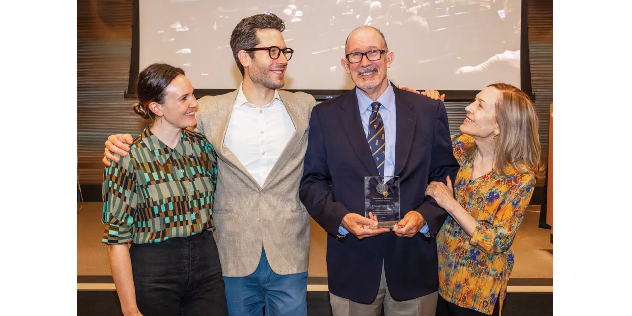
We sat down with Bill Dillon last summer after his official retirement, a hiking trip to Iceland and Norway, and just as he was beginning his next chapter as recall faculty. We asked him to describe himself in three words. He replied, “Oh, wow!” and then chose curious, devoted, and nature-lover, which aptly summarize a storied career in research and clinical practice, scores of creative and satisfying professional relationships, and a lifetime of outdoor adventures with his family and friends.
It was 1979 and Bill Dillon was an intern at Virginia Mason Hospital in Seattle. He felt uneasy in internal medicine – a specialty he chose largely because he loved all of medicine and struggled to decide. Acting on a friend’s recommendation, he flew to Salt Lake City to explore a radiology residency position at the University of Utah. Following an excellent interview, the department chair suggested a hike in the Wasatch Mountains – a perfect backdrop for a nature lover to make the “unbelievably fortunate” decision that transformed his career in medicine.
“Intellectually, radiology seemed much more interesting in terms of problem solving and it touched all of medicine,” said Dillon. “Without really knowing much about radiology, and certainly nothing about the future of radiology as it turned out, I decided to give it a go.”
Four Decades of Accomplishments
“I feel unbelievably fortunate to have been in this specialty,” said Dillon. “We have this incredible reach. We touch literally every patient that comes into the hospital, and several hundred thousand people per year who go to our outpatient facilities. Imaging, especially in an academic setting, is the cutting-edge of innovation.”
Dillon credits his mother for his curiosity and love of reading, early habits of mind they cultivated together at the Hayward Public Library in Alameda County where he grew up in a large, extended Irish and Croatian family. Propelled by the freedom to explore his academic and clinical interests, Dillon’s expertise evolved over more than 40 years. His began with head and neck radiology, specifically bringing MR imaging to the field. Then he focused on diagnosing and treating patients with brain tumors who frequently sought care at UCSF Health for these complex conditions. Most recently, he has become renowned for treating spine issues in patients with pain or CSF leaks.
Driven by Curiosity and a Mazda RX7
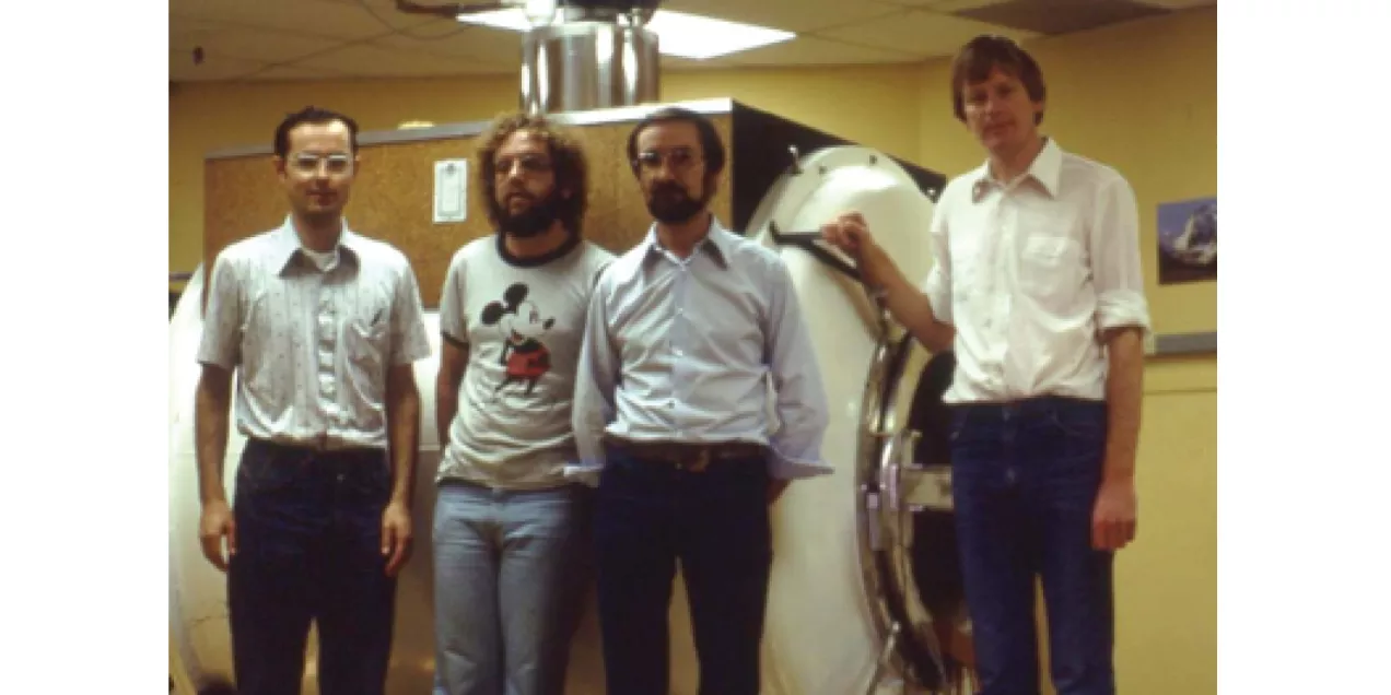
Over the course of Dillon’s career, technology has transformed medical imaging and Dillon acknowledges that he was often in the right place at the right time. Magnetic resonance imaging (MRI) was developed in the 1970s, culminating in the first MR scan of the human body in 1977, with MR scanners becoming commercially available in the 1980s. During his fellowship at the VA in 1982, Dillon’s curiosity led him to drive patients in his two-seater sportscar from the Veterans Affairs Medical Center to a UCSF research facility at Oyster Point that was home to one of the first MRI scanners on the West Coast. (With a smile, Dillon notes that this was before HIPAA!)
“I realized MR technology would make a huge impact. If I left academics, it would be several years before I’d experience it in private practice,” he said, crediting MR technology and his mentors with keeping him in an academic career. “That was a dramatic change to be able to image with that kind of fidelity without radiation.”
For around a decade early in his career, Dillon specialized in head and neck radiology, drawing on expertise from his time at the University of Utah, where he was trained and mentored by Anthony Mancuso, MD, who was among the small group of neuroradiologists who developed the subspecialty in the 1960s and 1970s.
Dillon began to use CT imaging for spinal imaging as early as 1983. Developed in the late 1960s and early 1970s, computed tomography (CT) revolutionized medical imaging. As the resolution and speed of CT imaging advanced – the first CT scanners took several minutes to acquire a single image, compared to today’s CT scanners that capture multiple images simultaneously in a fraction of a second – it became a vital tool in medical diagnostics, capable of providing detailed images of the body’s internal structures, including organs, blood vessels, and bones. In 1983, Dillon authored his first research paper on cranial CT. In 1984 his article, Magnetic Resonance Imaging of the Nasopharynx, was the first description of MR imaging in the field of head and neck radiology.
As CT technology accelerated, Dillon helped pioneer the replacement of invasive catheter angiography with CT angiography and perfusion CT, imaging techniques that use a contrast agent injected into the blood vessels to visualize and examine arteries, veins, and heart chambers to guide procedures and treat conditions such as stroke.
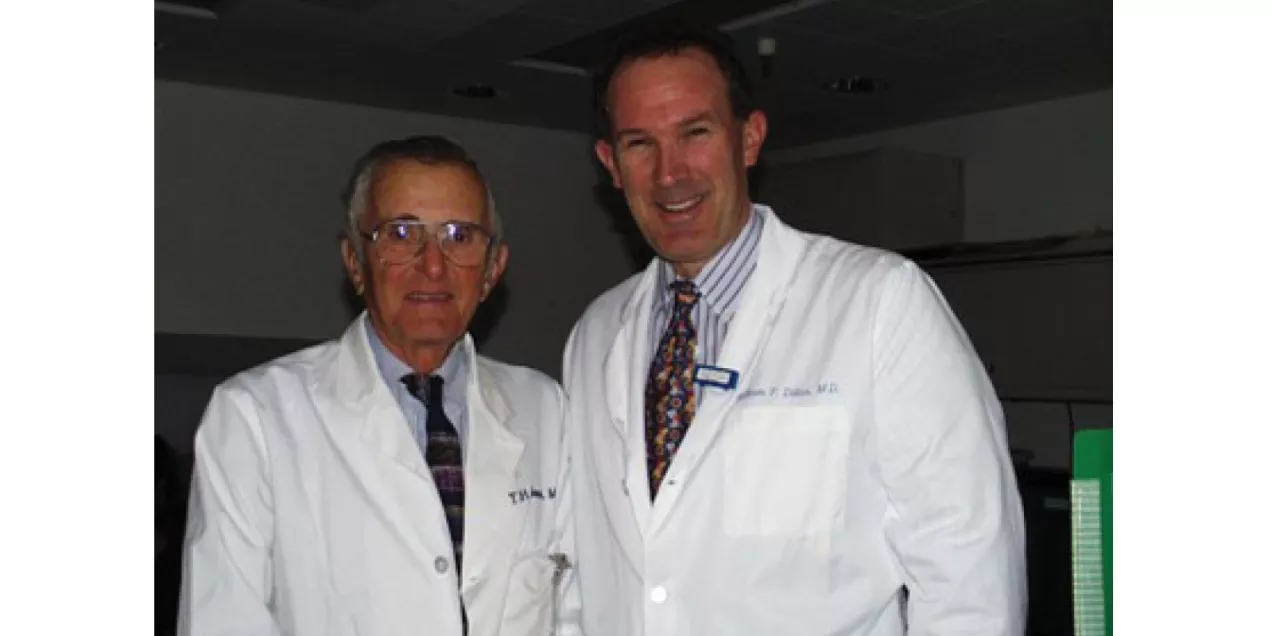
Reflecting on his service as chief of neuroradiology from 1992-2015 – the division was founded by his mentor T. Hans Newton – Dillon is quick to credit the group’s vibrant culture of innovation. Dillon noted that this leadership role prompted a natural shift to focus on complex brain tumors and related cancers, as significant numbers of patients were coming to UCSF seeking treatment for these conditions. For more than two decades, Dillon and his trainees partnered with research colleagues – including Sarah Nelson, PhD, and Dan Vigneron, PhD, who developed new tools for brain tumor imaging and characterization – to translate techniques developed in research labs into the clinic.
In the early 2000s, Dillon took a keen interested in spine imaging and pain management, including cerebrospinal fluid (CSF) leaks. Building upon his work in myelography and in partnership with Cynthia Chin, MD, Dillon developed new procedures that integrated high-field MRI and high-resolution CT for diagnosis and treatment of spine and peripheral nerve problems. By locating the source of pain and then delivering pain medication with pinpoint accuracy, UCSF has been at the forefront of improving patient outcomes for complex spinal pain, in large part due to Dillon’s leadership in establishing the UCSF Precision Spine and Peripheral Nerve Center at China Basin.
Roadmap to Success: Navigate the Bumps, Have Fun Along the Way
In a recent presentation to the American Society of Neuroradiology, Dillon described a bumpy road as he recognized the struggles of early career academic radiologists in the audience who feel pulled in many different directions, in an environment where change is constant. From applying for grants, to receiving funding, Dillon understands the energy and focus required to launch new research projects, teaching and mentoring, while also managing high volume clinical workloads and carving out time for family.
As for the future of radiology and imaging science, Dillon notes promising research on the horizon in mental health, theranostics, HIFU, and new image-guided procedures. He’s personally interested in global education for radiologists through Health4TheWorld, a non-profit organization that he co-founded with Bhavya Rehani, MD. Regarding challenges, Dillon flags artificial intelligence, specifically its ethical and value-added use in healthcare, and the fact that federal funding for research has been essentially flat, indexed to inflation, for two decades, which stifles innovation and academic progress.
When pressed for advice, Dillon suggested that there’s no substitute for the synergy that develops when radiologists and imaging scientists collaborate in person. He elaborated: “Focus on one or two areas of interest and become an expert, then define your goals, and stick to them. Get the advice of others who are further along in their academic careers – they’re here for that. Then network like crazy and accumulate lots of mentors and friends!”
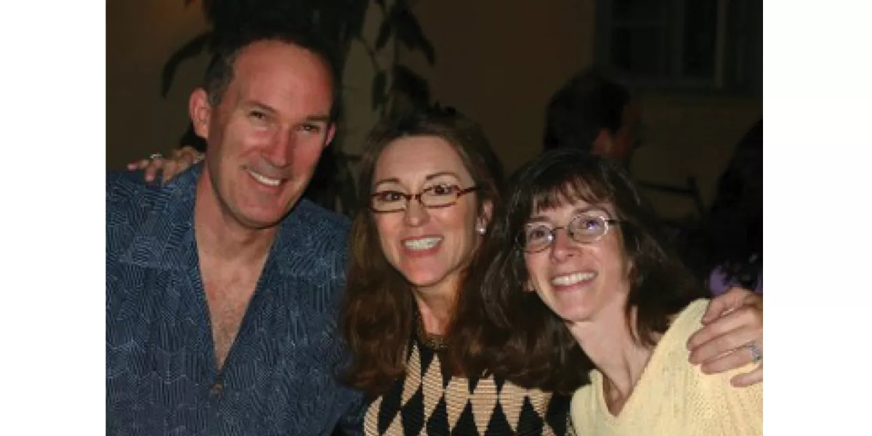
“You know your life goes by pretty quickly,” he said, reflecting on his career. “So, make sure that you’re having fun along the way!” He and his wife Irene, a former UCSF Radiology resident, enjoy travel and hiking in the mountains and have completed several classic hikes including the Tour d Mont Blanc, the Haut Route, and most recently hikes in Sardinia and Corsica.
William Dillon, MD, is professor emeritus and was the Elizabeth A. Guillaumin Professor of Radiology from 2004 – 2024. He joined the UCSF Radiology faculty in 1983. Dr. Dillon received his medical degree from UCLA in 1978 and completed his residency at the University of Utah in 1982, followed by a neuroradiology fellowship at UCSF in 1982-3. He is a co-founder of a non-profit, Health4theWorld.org.
Honors
- Francis A. Sooy, MD, teaching award for Clinical Excellence, UCSF Otolaryngology, 1993
- Gold Medal, American Society of Head and Neck Radiology, 2008
- J. Elliott Royer award for contributions to clinical neurology, UCSF Psychiatry, 2011
- Outstanding Researcher, American Society of Neuroradiology, 2014
- Gold Medal, American Society of Neuroradiology, 2015
- Outstanding Faculty Mentoring, UCSF Radiology and Biomedical Imaging, 2016
- Gold Medal American Society of Spine Radiology, 2025
Leadership
- Chief, Neuroradiology, San Francisco VA Medical Center, 1983-86
- Chief, Neuroradiology, UCSF, 1992-2015
- President, ASHNR, 1993
- President, ASNR, 2000
- Executive Medical Director for Ambulatory Imaging, UCSF Health, 2018-25
- Executive Vice Chair, UCSF Radiology and Biomedical Imaging, 2007-24
- Dr. Dillon was a senior editor for the American Journal of Neuroradiology (1998-2011) and has been a co-investigator in 25 funded projects and 10 clinical trials. He has published 300+ articles and 30+ book chapters.