
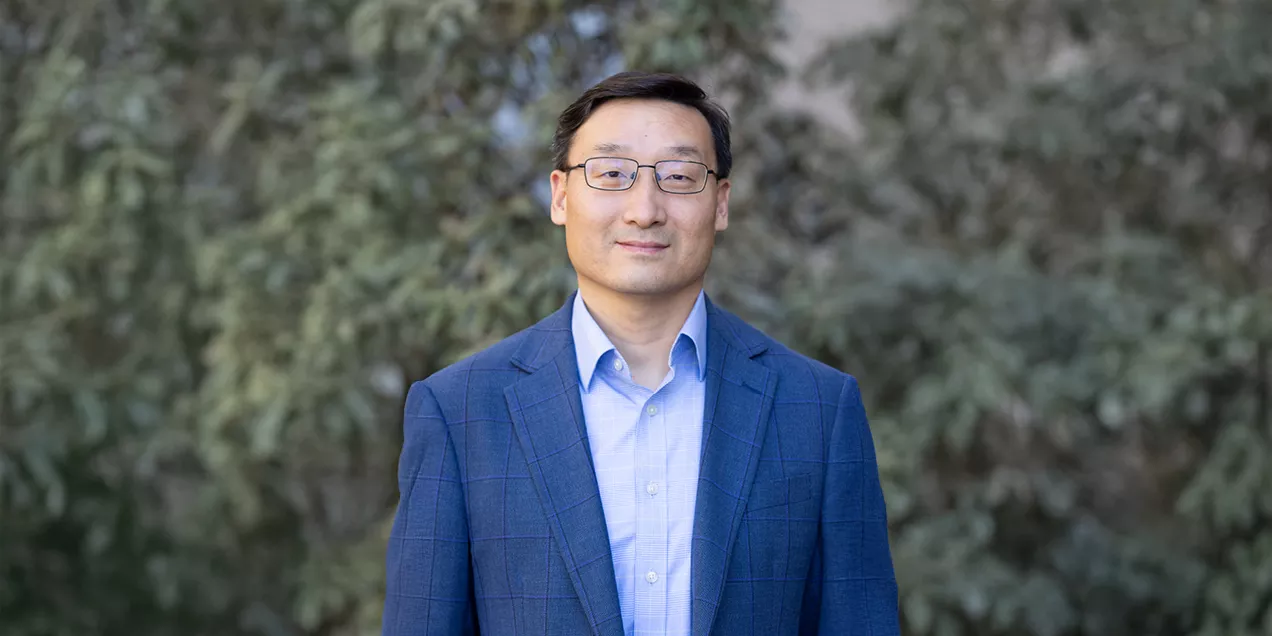
For nearly two decades, imaging scientist Zhaoyang Fan, PhD, has developed and refined MRI techniques that reveal how blood vessel walls change over time. A recent addition to the UCSF faculty, Fan is professor in residence and co-chief of Cardiac and Vascular Imaging Research, an imaging science group that spans radiology, neurology, cardiology, and engineering.
Black Blood Imaging
Most MRI scans capture a single moment. For patients with stroke, aneurysm, or hypertension who receive multiple MRI scans over many months, physical changes in vessel walls that develop in the intervals between scans provide valuable data for clinical decision-making.
Fan has focused much of his research on vessel wall – or “black blood” – imaging, an MRI technique that visualizes artery walls instead of the blood flowing through them. Black-blood imaging allows clinicians to identify high-risk atherosclerotic plaques that can lead to stroke and monitor how those plaques respond to treatment over time. With his background in technical development, Fan’s research focuses on making black blood imaging techniques more robust.
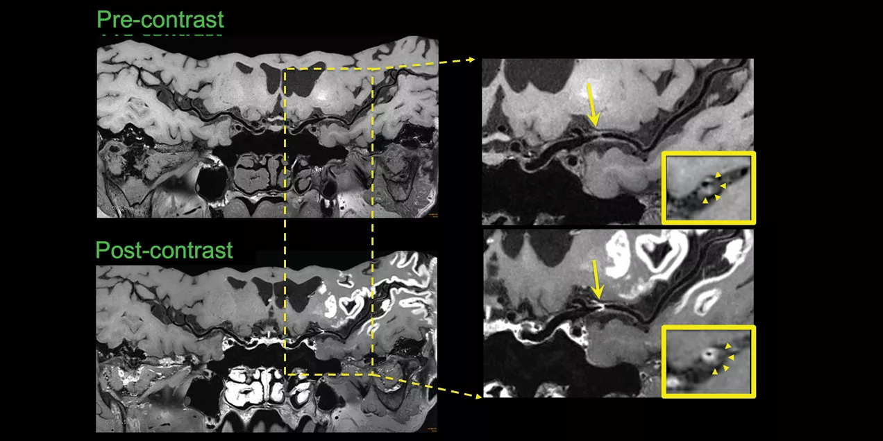
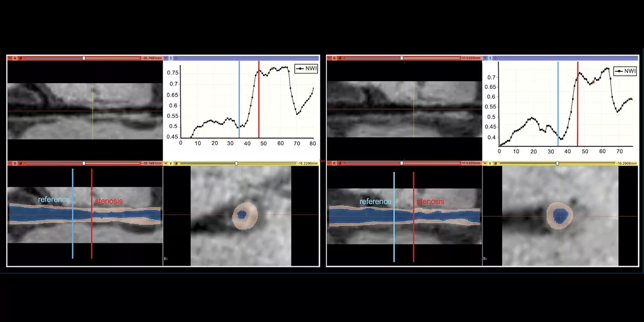
His laboratory has refined nearly every step of the imaging process. They have developed sequences to suppress the blood signal completely while implementing new acquisition methods that reduce scan times to less than four minutes. Together, this makes examinations more comfortable for patients while reducing motion artifacts. Automated analysis software converts subjective image interpretation into quantitative measurements that radiologists can track over time.
"We're not just saying something looks brighter or thicker," Fan said. "Now we can provide real numbers. The change between two time points is more important than the baseline image. That's what tells you whether the patient is responding to medical therapy."
For physicians treating stroke patients, that information has practical consequences. The measurements help physicians identify patients responding to therapy and those who may need more aggressive treatment before another stroke occurs.
Carotid atherosclerotic plaque is a major cause of stroke in Fan’s home country of China and other East Asian countries. Fan has spent decades on these imaging techniques because he wanted to make a difference in the clinical management of that patient population, to produce novel techniques that can identify high-risk lesions and make effective treatment decisions to avoid recurrence. He observes that, “As a scientist, I try to be responsible, be open to collaboration, and to make high-impact research that serves our society."
Diagnosing Stroke Etiology Faster
Fan collaborates with neurologists to more accurately diagnose stroke etiology with new combined MRI protocols for the head, neck, and chest. When a patient is admitted to the hospital with an ischemic stroke event, neurologists want to identify the cause – typically a thrombus (blood clot) that forms at the site of a fatty plaque in a blood vessel that supplies the brain or an embolism (blood clot) that forms in the heart or arteries of the chest or neck. Physicians treat each type of blood clot differently, and timely identification helps reduce the risk of recurrent ischemic stroke, which is often more severe than the first event.
"We hope to give our physicians powerful imaging tools to make quicker and more accurate diagnoses," Fan said. "So they know how to treat and manage their patients."
Fan aims to develop a quick imaging technique integrating two scans into one MRI imaging protocol, looking at the heart, the aorta, and the carotid and intracranial vessels. Acute ischemic stroke patients all receive a brain MRI exam after admission, and this additional protocol could inspect all suspicious etiologies in the major stations in less than 10 minutes. He recently received NIH funding for this study, a collaboration with the Department of Neurology and serving patients at Zuckerberg San Francisco General Hospital (ZSFG).
Building on Collaboration
Long before arriving at UCSF, Fan had established research partnerships with David Saloner, PhD, and Kambiz Nael, MD, developing new MRI methods for intracranial vascular disease. For Fan, collaboration is more than sharing expertise. He believes successful partnerships are built on mutual benefit, with each investigator contributing distinct strengths.
One of the biggest turning points in his career came during the COVID-19 pandemic. While laboratories became quieter, virtual meetings made it easier to connect with investigators across the country, leading to new collaborations and multi-institutional NIH grant applications.
When Fan looked for outside collaborators on his new black blood imaging technique, Saloner, a friend and colleague for more than a decade, was at the top of his list.
Consistency and partnership allow these discoveries to advance, Fan observes, reflecting that, "You have to really work in a field for a long time. Scientific advances rarely come from a single discovery. They emerge through years of refinement, sustained collaboration, and careful observation.”