
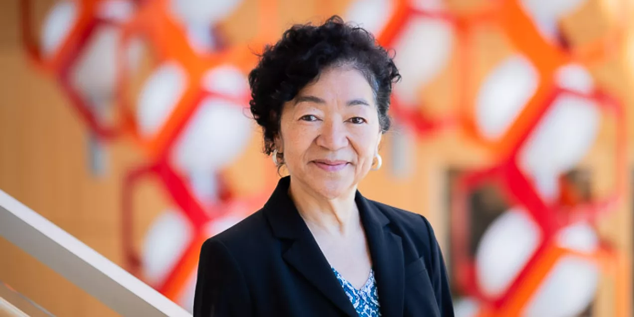
Nola Hylton, PhD, Earns National Academy of Medicine Recognition
One of the highest honors in the field of health and medicine, the National Academy of Medicine recognized Nola Hylton’s work in the early development of MRI imaging for the detection, diagnosis and staging of breast cancer, and for improving the health of millions of women globally.
As director of UCSF’s Breast Imaging Research Group, Hylton’s laboratory collaborates with radiologists, surgeons, oncologists and researchers around the world to optimize MRI techniques for breast cancer patients.
In a recent interview, Hylton answered a few questions about her career –
What inspired your work in MRI technology?
During my PhD training, MRI was moving from a lab tool into the clinical setting. My Stanford advisor Al Macovski was going on a year-long sabbatical to Europe, but I needed to get traction on my research, so he arranged for me to work in this incredible UCSF lab at Oyster Point where they were building an MRI system, all the electronics, the software, everything. I was lucky to be part of that, and it helped me understand fundamentally how the machine produced pictures. I would sign-up for scanner time on a Wednesday, and I had the machine to myself all day. Unheard of now!
The practicality here is that the major manufacturers of MRI equipment saw a market, and they started to build radio frequency coils, which are designed anatomically for the head, leg, ankles. The breast was an obvious focus because of the prevalence of breast cancer. But the design of the coil was challenging.
When breast coils did become available our department chair at the time, Dr. Alex Margulis, said “We’re getting a breast coil. You should start doing this work!”
How did you become a breast cancer researcher?
My training up to this point was in male dominated fields – chemical engineering at MIT and applied physics at Stanford. When I moved to breast cancer research, the patients and most of the treating physicians were women. It was remarkable to be in an environment where a technology that I really enjoyed was suddenly much more relevant to my own life.
Within a few years, there was an international effort to develop MRI technology. In 1990, the NIH established the Office of Research on Women’s Health and Alex Margulis sent me to one of their meetings. When I look back on it, there were luminaries from around the world collaborating on a multicenter trial. Had I not been exposed to these investigators so early in my career – many of whom are my colleagues and friends decades later – I don’t know that I would be working in clinical trials so strongly now.
The group was tackling questions like What should the equipment be? What should be the standards for acquiring data, for measuring, for interpreting? We are all highly opinionated people who debate and disagree. But I don’t really see that as a nonstarter because, in fact, we all want this technology to be applicable and we all care about patients and doing good science.
What are your thoughts on the future of MRI in breast imaging?
While my area is imaging and MRI, I think of myself first as a breast cancer researcher interested in breast cancer risk factors, who develops breast cancer, and the likelihood that a new technology will be meaningful in the setting of breast cancer.
There are so many clever things we can do with MRI. It’s attractive from the physics point of view. But I’m painfully aware of the fact that what we do is not available to most women with breast cancer who don’t have access to academic centers or insurance coverage for MRIs during their oncology care.
We might make breast MRI more deliverable and miniature, and probably use AI to transform the technology into something much more feasible and inexpensive – there are portable, low-cost machines now. It sounds futuristic, but I see potential for handhelds with the ability to scan using an MRI phenomenon. I don’t think we’re tied to MRI, I think we’re tied to a technology that tells us about the amount of response a tumor is having. Many options are available now -- liquid biopsies, contrast-enhanced mammography, blood markers.
As a field, we must figure out what’s the best way to monitor treatment. You don’t always need a whole MRI exam. And then there are times when we really do need the exquisite picture of what’s there before surgery, where things are, and what needs to be taken out.
NAM recognizes individuals who have demonstrated outstanding professional achievements and commitment to service in the medical sciences, health care and public health. The academy elects no more than 100 members a year.