

Resurgence of SPECT and SPECT/CT for Quantitative Imaging of Radiotheranostics
To ensure radiopharmaceutical therapies such as 131I-NaI for thyroid cancer, 90Y-microspheres for liver cancer, 177Lu-DOTATATE for neuroendocrine tumors, and 177Lu-PSMA-617 for prostate cancer, are both safe and effective, we must move beyond a standard-dose approach and embrace individualized dosimetry. Each patient's body processes these targeted drugs differently. Therefore, an important need exists for quantitative imaging that can precisely measure how much radiation is delivered to tumors as well as normal tissues and organs. This allows us to modulate treatments, maximizing the therapeutic efficacy while minimizing potential toxicity from radiation. At our UCSF Physics Research Laboratory (PRL), our focus has been on developing and refining the quantitative imaging technologies, which coincides with the resurgence of SPECT/CT in the era of radiotheranostics, an important step toward precision oncology.
Rationale for quantitative SPECT/CT
A major achievement of PRL has been the advancement of quantitative SPECT/CT. Historically, nuclear medicine imaging was primarily qualitative, showing where a radiopharmaceutical accumulated but not accurately how much. UCSF PRL not only pioneered the invention of SPECT/CT (led by the late Bruce Hasegawa and his team then) but has pushed several important technological advances in many clinical and preclinical applications, all toward making SPECT truly quantitative, i.e., physically accurate in its measurement.
The core principle of quantitative SPECT using SPECT/CT is to convert the raw image data, the detected photon counts in each voxel, into a true, physical unit of radioactivity concentration, such as Bq/ml. To achieve this, we must correct for all known major physical perturbation terms that degrade the image quantitativeness, including: a) photon attenuation, b) photon scatter, and c) collimator-detector response at minimum.
Using the anatomical map from the integrated CT scan, we can accurately model and correct for this attenuation on a patient-specific basis. The scatter is incorporated by combining high energy resolution detectors such as cadmium zinc telluride (CZT) and energy or geometry-based scatter count subtraction. Collimator-detector response is corrected by modeling and measurement, a technique basically mathematically identical to deblurring of positron emission tomography (PET) based on point-spread-function (PSF) responses by detectors to measured blurring.
By systematically addressing these factors, we can generate SPECT/CT images that are physically accurate 3D maps of radioactivity distribution. This quantitative information is the essential input for radiation dosimetry, allowing us to calculate the absorbed radiation dose to specific tumors and normal healthy organs like the kidneys and bone marrow. Without quantitative SPECT imaging techniques that our lab has pioneered, the radiation dosimetry of those very commonly practiced radiopharmaceutical therapies is not even feasible.
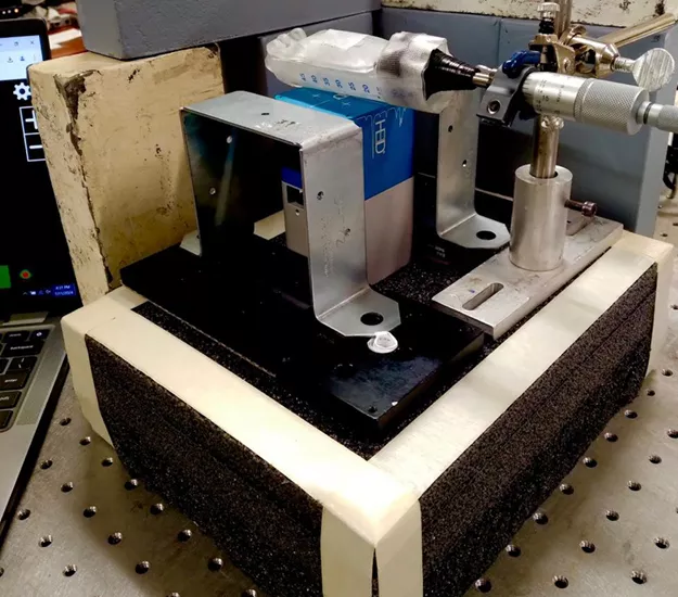
Advancements in detector technology: CZT-SPECT
Building on this foundation, we have also focused on next-generation hardware. A key area of our development is the application of CZT-based SPECT systems (Figure 1). Unlike traditional radiation detectors, CZT provides superior energy resolution by directly converting the high-energy gamma-ray interactions to electrical charges. This technological advance allows us to better separate scattered photons from primary photons, making SPECT basically more quantitative and visually improved image quality by this hardware approach.
The challenge of imaging alpha emitters like Actinium-225
We are also tackling one of the significant challenges in targeted alpha therapy: imaging Actinium-225. With our understanding that there is no image quality if there are not enough photons detected, our approaches have heavily focused on improving photon detection efficiencies by orders of magnitude if possible. As a potent alpha-emitter, 225Ac is highly effective at killing cancer cells when delivered precisely on target, but its direct gamma emissions are very low, making them very difficult to image accurately.
Our lab investigates novel imaging strategies that use the gamma emissions from 225Ac's daughter nuclides. With new or repurposed technologies used for other purposes, our goal is to achieve reliable in vivo quantitative imaging of 225Ac-based radiopharmaceuticals at clinically acceptable radioactivity levels that are typically extremely low. This work is essential for confirming that the targeted alpha therapy delivery is on target, and how much off-target toxicity could be expected from the therapy so that both development of new 225Ac-based therapies and improving precision oncology efforts of clinical-stage 225Ac-based therapies can be advanced.