
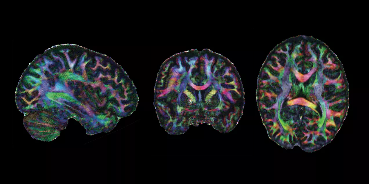
Next Gen 7T MRI Allows Neuroimaging with Unprecedented Precision
Conventional MRI scanners for clinical use generate images using magnets whose strength is measured at 1.5 Tesla (T) or 3T. At a limited number of locations across the world, clinicians and researchers have access to MRI scanners with much more powerful magnets operating at 7T. With the several dozen 7T machines located in the United States, imaging scientists can gain much higher resolution images, examining the tiny details that make up the architecture of the brain and body. NexGen 7T 0.9 mm isotropic diffusion tensor images, shown above, demonstrate the impressive anatomical quality and detail that can be visualized throughout the brain. However, there is still a ceiling to that resolution, as even with the most optimal scan parameters, features smaller than one to two millimeters vanish into the abstraction of the voxel, a three-dimensional pixel.
In December 2023, an international team of scientists including UCSF faculty member An (Joseph) Vu, PhD, broke that ceiling. Led by UC Berkeley professor and president of Advanced MRI Technologies, David Feinberg, PhD, MD, the team is a multi-institution collaboration with scientists from UCSF including Drs. An (Joseph) Vu and Pratik Mukherjee, UC Berkeley, Harvard, Siemens Healthineers (Erlangen, Germany), Advanced MRI Technologies (Sebastopol, CA), and MR CoilTech LTD (Glasgow, UK). Together, they constructed a next generation ultra-high resolution 7T MRI scanner, which achieves up to 10-fold increase in resolution over the current 7T standard, which is 50 times more detail than the hospital standard 3T scanners. With this new tool, functional images can now be captured with a voxel size
of less than half a millimeter. This allows scientists to, for the first time, image functional clusters of neurons across the entire brain, organized in cortical cell layers and cortical columns, opening up the study of a new realm of meso-scale local neurocircuitry.
Thanks to additional funding obtained by Drs. Vu, Feinberg, and Alexander Beckett, the NexGen 7T is now available to scientists across the Weill Neurohub consortium of UCSF, UC Berkeley, and the University of Washington, and as an international resource through the NIH BRAIN Initiative. The NexGen 7T MRI scanner is described in a Nature Methods article “Next-generation MRI scanner designed for ultra-high-resolution human brain imaging at 7 Tesla.”1
Over the years, the San Francisco Bay Area has had as many as four 7T scanners, each dedicated to state-of-the-art imaging research at UCSF Mission Bay, the SF VA Medical Center, UC Berkeley, and Stanford. Since 2017, FDA-approved 7T scanners have become available, fueling a growing push to make this technology more readily accessible in clinical environments.
Although the NexGen 7T is not yet FDA approved, it has achieved remarkable breakthroughs in fast, high-resolution neuroimaging by virtue of several key advancements: an extremely powerful head gradient system, the first 128 channel receiver systems integrated into a 7T scanner, and several universally optimized RF pulse sequence protocol.

The new head-only magnetic gradient coil design that is an order of magnitude more powerful than those in commercially available 7T scanners. This advancement in gradient performance was achieved with a novel 3-layer wire winding design instead of only two layers. The faster and stronger the magnetic gradients are, the faster the MRI data can be encoded, fighting the clock of T2 signal decay and blurring. There were a lot of physics challenges to achieve such strong and fast gradients. In addition to the need to minimize peripheral nerve stimulation effects, the mechanical forces interacting with the field and the sound pressure levels both increase as the main magnetic field gets stronger. These challenges were detailed in “Acoustic noise reduction in the NexGen 7 T scanner.”2 For all these reasons, the scanner needed to be designed at a system-wide level, factoring in RF coil design, gradient coil design, and magnet design.
Another breakthrough came via the development of 64 channel and 96 channel receiver arrays coupled with the 128-channel receiver system, vastly improving on the standard 32 channel system in terms of SNR and the ability to accelerete the imaging. As the number of receiver channels increases, the size of the individual coil loops in the head coils become smaller, which in turn provides higher sensitivity and improved ability to accelerate data acquisition for fast, ultra-high resolution functional and structural MRI.
The improvements are not only related to hardware, as RF pulse sequence design has played a great role in these milestone achievements. In collaboration with Dr. Nicolas Boulant (CEA, NeuroSpin, France), the team has implemented precisely pre-calibrated universal pulses which can produce structural images of exquisite quality on almost any subject you place inside the scanner without the need for lengthy pre-scans or subject-specific calibrations. Furthermore, in two collaborations with Dr. Renzo Huber (NIMH, NIH) and with Dr. Suhyung Park (Chonnam National University, S. Korea), improvements in functional imaging pulse sequences more precisely identify neuronal activity in cortical layers, and similar resolution gains in collaboration by achieving unprecedented isotropic resolution in the 0.35mm – 0.6mm range.
Feinberg points out, “The NexGen 7T scanner achieves greatly improved precision in diffusion imaging of axonal fiber tracks from the cumulative gains of high signal from 7T and the much stronger gradient encoding, now possible. Secondly, the scanner’s ability to achieve mesoscale functional imaging at depths in the cortex rather than averaging across the cortex provides more precise information to take new directions in neurocircuitry studies of different neurological disorders including depression, chronic pain, localization in epilepsy and revealing the underpinnings of many cognitive disorders.”
Vu is excited about the new avenues this next-gen 7T scanner will open up, explaining that “Traditionally, the RF pulse sequence optimization is a very involved process done on a per subject basis. One had to acquire calibration scans, model the head, and calculate how best to excite the whole brain. Not every 7T site has the time, expertise, and capability for such an optimized scan protocol. However, with the new universal pulses pre-calibrated technique the images come out very nice on any subject, right out of the box. In the past, some clinicians and collaborators have been hesitant to go to 7T because such technology was not readily available. But with these new NexGen 7T technologies, it removes the hesitancy bottleneck for wide-spread adoption into clinical neuroimaging and research. It is a game changer!”
References
1) Feinberg, D.A., Beckett, A.J.S., Vu, A.T. et al. Next-generation MRI scanner designed for ultra-high-resolution human brain imaging at 7 Tesla. Nat Methods 20, 2048–2057 (2023). https://doi.org/10.1038/s41592-023-02068-7
2) Boulant N, Ma S, Walker E, et al. Acoustic noise reduction in the NexGen 7 T scanner. Magn Reson Med. 2024; 92: 2261-2270. doi: 10.1002/mrm.30211