Neuropsychiatry Research Roundup
UCSF Radiology and Biomedical Imaging clinicians and imaging scientists reveal new facets of brain-behavior relationships in emotion, obsessive-compulsive disorder (OCD), autism, epilepsy, and neuropsychiatric illness.
Mapping Joy in the Brain with Advanced fMRI Techniques
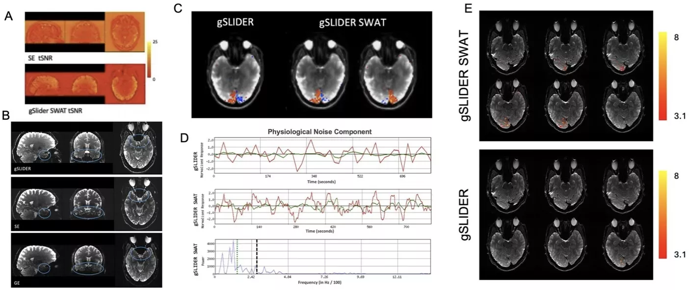
What happens when the brain experiences joy? An Vu, PhD, and collaborators introduce a novel fMRI method, gSLIDER-SWAT, designed for high spatial-temporal resolution imaging of the brain on widely accessible 3T MRI scanners. This technique captures high-resolution brain activity associated with joy, pinpointing key regions, such as the basolateral amygdala, reward centers, and visual cortex. Their work offers a clearer view into emotional networks that are often challenging to detect with standard imaging, opening doors to refined investigations of positive emotion processing.
Deep Brain Stimulation’s Impact on OCD’s Neural Networks
Deep brain stimulation (DBS) targeting the anterior limb of the internal capsule can bring relief to some individuals with severe, treatment-resistant OCD. Using functional MRI during stimulation on and off cycles, Melanie Morrison, PhD, and collaborators observed that effective therapy reduced activity in regions of the default mode network, such as the orbitofrontal and dorsomedial prefrontal cortex, along with the subthalamic nuclei. Structural connectivity findings indicate that this suppression of activity may result from disrupted communication along white matter pathways tied to the brain target stimulated by DBS.
Gene-Brain-Behavior Links in Autism via Machine Learning
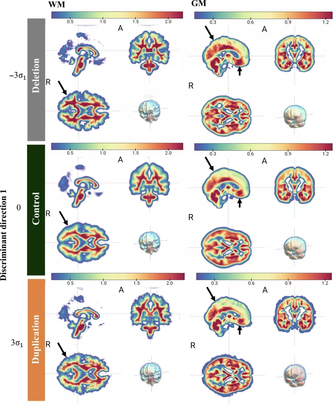
Using 3D transport-based morphometry, Pratik Mukherjee, MD, PhD, and collaborators show how genetic variations at the 16p11.2 locus shape brain structure and behavior in autism. Analyzing data from the Simons Variation in Individuals Project, they identified two brain endophenotypes predictive of brain alterations connected to articulation and intelligence differences, illustrating the power of genetics-first imaging to uncover neurodevelopmental diversity.
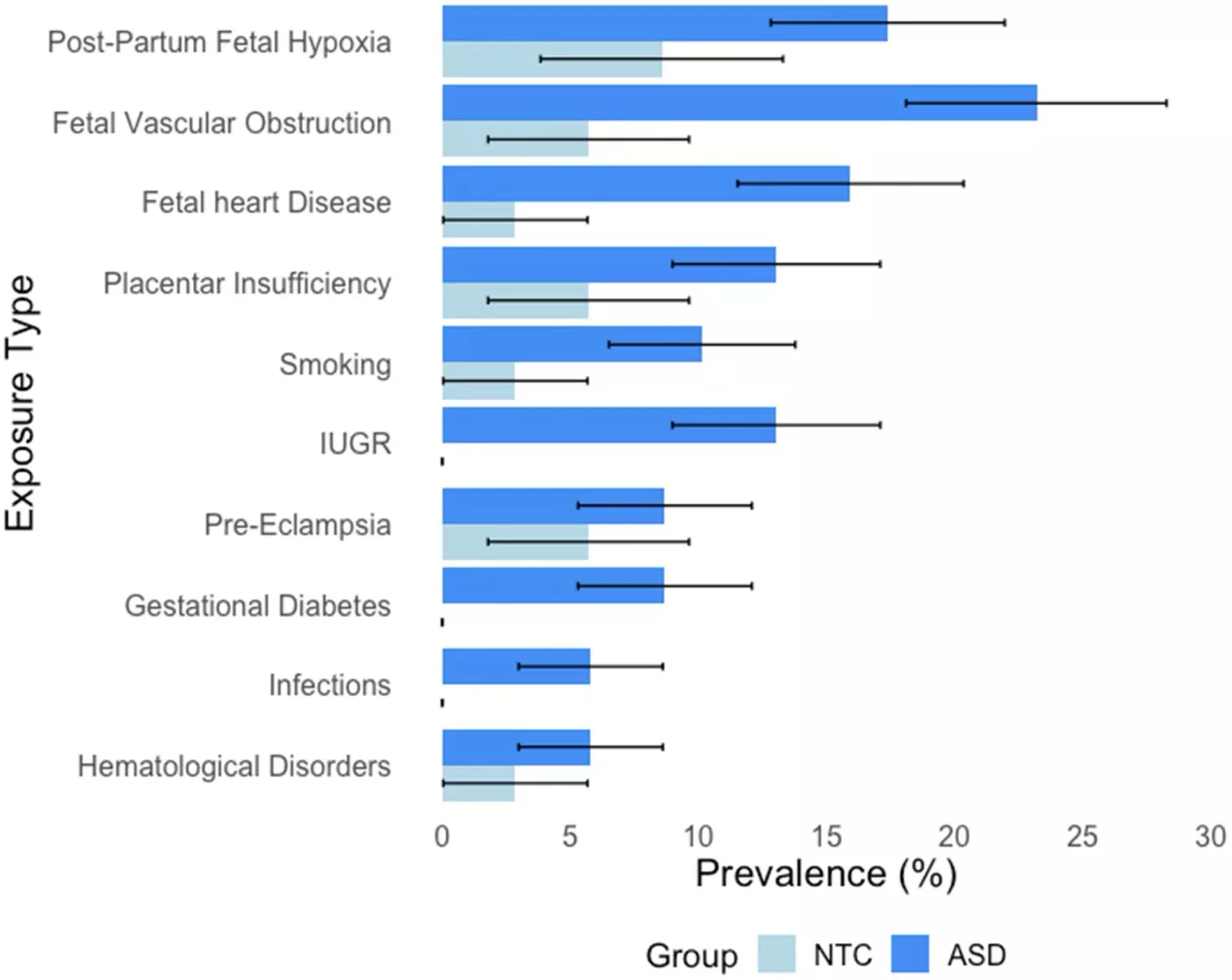
Prenatal Oxygen Deprivation Influences Autism
Youth with autism were more likely than neurotypical peers to have experienced prenatal hypoxic risk conditions, according to this study of 104 participants. This study by the Yan Li lab found higher rates of oxygen deprivation exposure in autistic participants, linked to enlarged third ventricle volumes and more severe sensory and sleep disturbances. These results suggest that oxygen deprivation before birth may alter brain development in ways that intensify certain autism traits, highlighting the importance of targeted prenatal care and early interventions to reduce these risks.
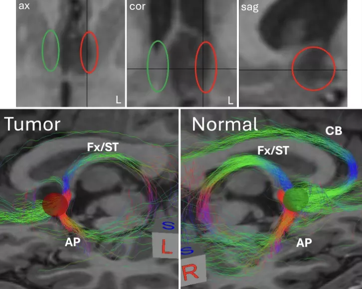
Tumor Resection Quickly Resolves Depression and Epilepsy
In this illustrative case by Pierre Nedelec, Leo Sugrue, MD, PhD, and collaborators, a young patient’s depression and seizures vanished rapidly after surgery to remove a low-grade tumor from the septal region, a limbic hub tied to mood regulation and reward processing. The outcome suggests that structural lesions in this area can drive both mood disorders and epilepsy, offering fresh insights into how targeted interventions might address complex neuropsychiatric conditions.
AI Reveals Pathology and Psychosocial Drivers in Back Pain Opioids
Factors influencing how we treat patients with chronic pain extend far beyond the presenting complaint. In this study, Sharmila Majumdar, PhD, led an AI-driven analysis on the importance of demographic and psychosocial factors. Findings highlight context-dependent prescribing behaviors, supporting a more nuanced, patient-specific approach.
The association between female sex and stronger medication prescriptions raises questions about potential gender biases in pain management, warranting further investigation. Current and former smokers were more likely to receive opioids but not NSAIDs although the predictive power was weak. Stronger prescription usage was associated with anxiety, depression, and between being partnered rather than single, suggesting clinicians consider psychosocial distress when prescribing opioids. These findings clarify how multiple patient dimensions interact to shape complex prescribing decisions for chronic pain, by quantifying the relative influence of various psychosocial and pathological factors.
This study offers healthcare institutions an actionable framework to systematically evaluate their management practices and identify opportunities for improved care for patients with chronic pain.
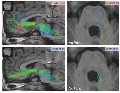
Understanding Bladder Side Effects of DBS for Depression
Deep brain stimulation (DBS) has shown promise for the treatment of major depressive disorder. However, DBS can lead to side-effects including bladder spasms. A case report by Leo Sugrue, MD, PhD, and Pierre Nedelec, MS, MTM, found that bladder spasms in a patient being successfully treated for depression with closed-loop DBS targeting the ventral capsule were likely elicited by activation of descending cortical pathways involved in regulating the pontine micturition center in the brainstem.
The patient’s bladder spasms started approximately 37 days after initiation of stable stimulation and detector settings and continued for over a month before the patient reported the side effect. By testing different stimulation amplitudes and contacts in the laboratory setting and modeling the specific connections being recruited by settings that elicited bladder spasms the team was able to identify the brain circuit causing the spasms and adjust the stimulation parameters to avoid activating this circuit while maintaining a therapeutic antidepressive effect.
This case provides insight into the neural circuit underlying bladder control and function and demonstrates the importance of understanding the complex connections of regions targeted during DBS in order to achieve therapeutic benefit while avoiding off-target side effects.
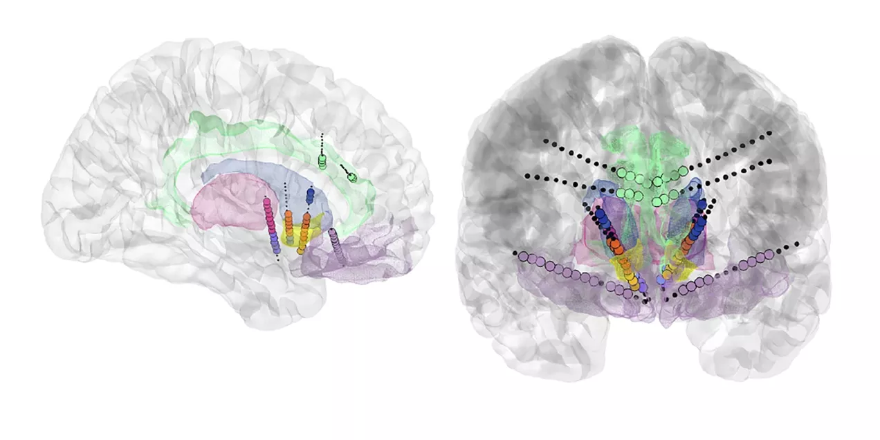
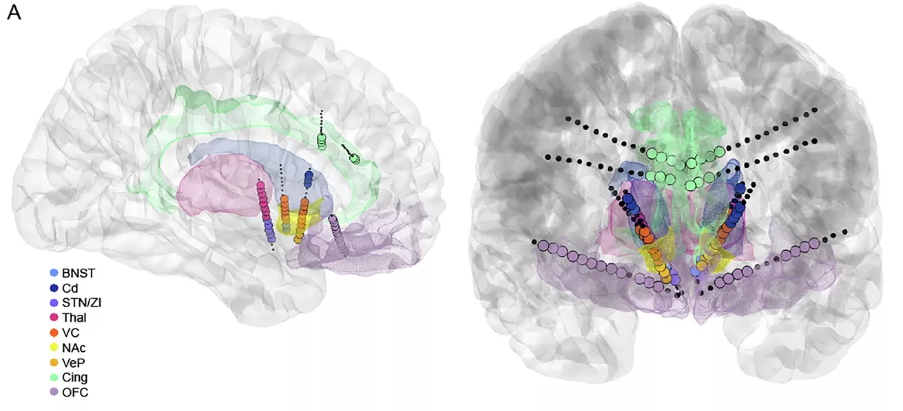
Brain Mapping Identifies DBS Targets to Treat OCD
Deep brain stimulation has been used to treat severe, refractory obsessive-compulsive disorder (OCD) with variable outcomes. To better map treatment areas, Leo Sugrue, MD, PhD, helped researchers implant electrodes across the implicated cortico-striato-thalamo-cortical circuit in a patient with severe refractory OCD. In this clinical trial, the research team performed extensive stimulation mapping using these electrodes during a multi-day inpatient stay to identify personalized therapeutic targets and characterize their connectivity and downstream circuit effects.
Two targets within the right ventral capsule were found to acutely reduce OCD symptoms and were both characterized by strong electrophysiological and structural connectivity to the orbitofrontal cortex. These sites were then implanted for long term DBS treatment. Combined stimulation of these targets led to a rapid therapeutic response, providing the first proof-of-concept that invasive brain mapping in combination with MRI assessments of brain structural connectivity can guide a novel personalized, multi-site neuromodulation to treat OCD.
The Cocktail Party Problem: Listening Difficulties with Background Noise
Understanding someone in a noisy room is always hard, but for some people it is almost impossible. Srikantan Nagarajan, PhD, and collaborators used MEG in a longitudinal cohort study to investigate the neural mechanisms underlying speech-in-speech listening in adolescents with normal hearing but listening difficulties (LiD). These children can experience developmental delays because they have difficulty understanding speech in complex listening environments.
Participants performed a task involving target speech presented alone or alongside competitor speech streams differing in talker and spatial cues. The team found that LiD, also known as the “cocktail-party problem,” arises primarily from selective impairments in processing attended speech – the ability to focus on one sound amid background noise – rather than heightened processing of competing sounds or general auditory disengagement. This assessment matched the caregiver-reported listening difficulties, validating this neurophysiological measure as a meaningful index of real-world listening challenges. The neural speech tracking index used was developed at UCSF.

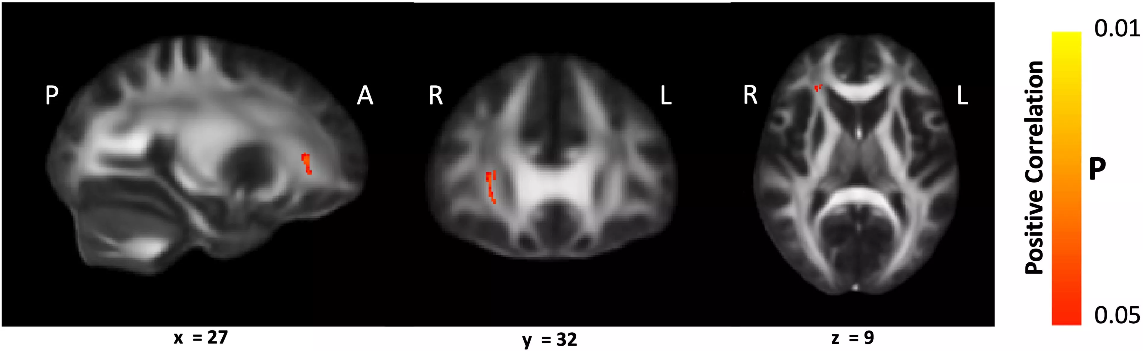
Cognitive Rehabilitation Influences White Matter Integrity in Brain Injury
Pratik Mukherjee, MD, PhD, led a team to understand the neural mechanism underlying attention impairment, one of the most common, debilitating, and persistent consequences of traumatic brain injury (TBI).
Goal-Oriented Attentional Self-Regulation (GOALS) is a cognitive rehabilitation training program that targets executive control functions in participants by applying mindfulness-based attention regulation and goal management strategies. The study found there was significantly better white matter microstructural integrity in left and right anterior corona radiata (ACR) in the GOALS group compared with the control group. They found a significant correlation between sustained attention ability of GOALS participants and white matter integrity of their right ACR pre- and post-training and that improvement was the result of increased neurite density and decreased fiber orientation dispersion within this tract.
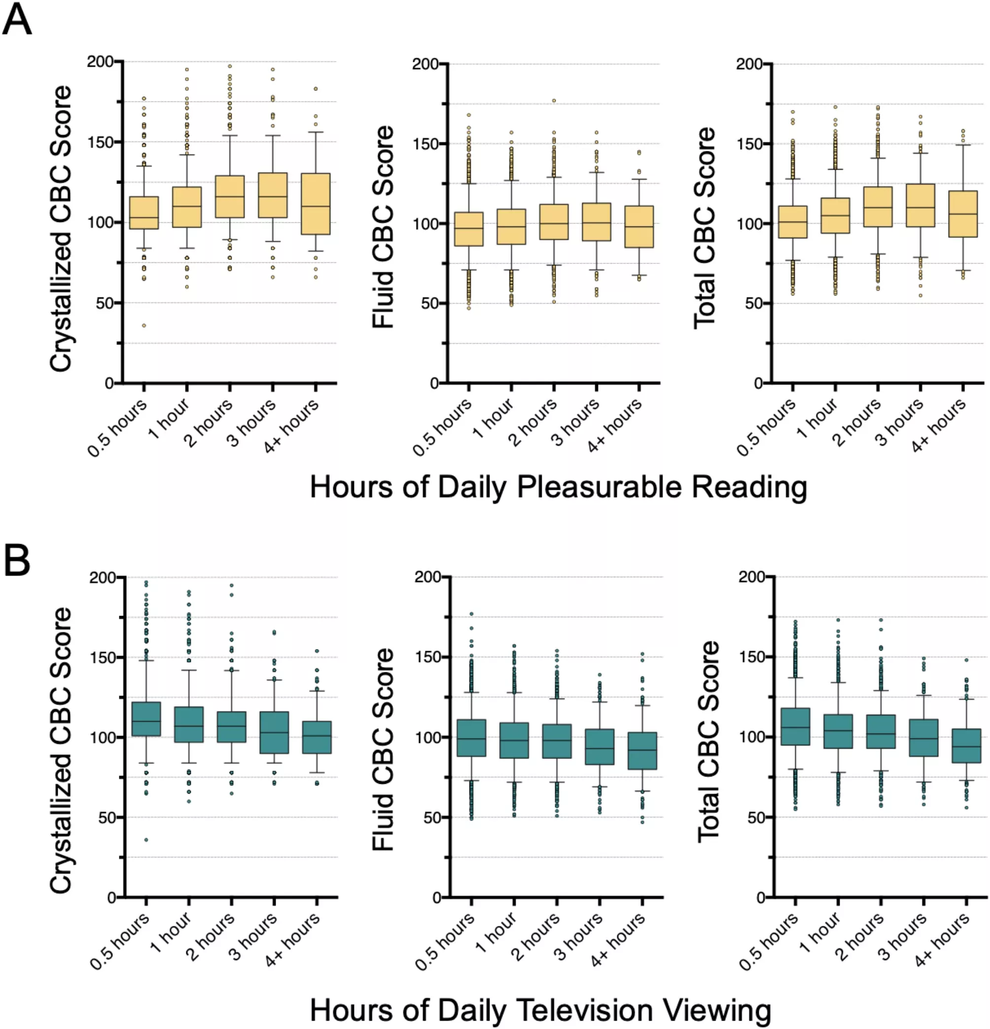
Reading and TV Shape the Growing Brain
By analyzing brain scans (MRI) and cognitive tests from over 8,000 adolescents in the US, co-first authors Andreas Rauschecker, MD, PhD, and Pierre Nedelec, MSc, with senior author Leo Sugrue, investigated how reading and television habits are linked to brain structure and cognitive abilities. Adolescents who reported spending more time reading for pleasure performed better on cognitive tests and showed an increase in the size of specific areas of the brain's outer layer (cortex). Conversely, more TV time was linked to slightly lower cognitive performance and subtle decreases in the size of some brain regions.