
AI Improves Meningioma Measurement
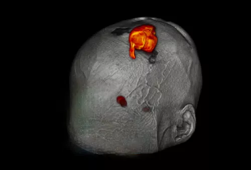
Meningioma is the most common type of brain tumor, and it is usually slow-growing which can make subtle changes difficult to detect. A new AI-enhanced tool developed by data scientist Pierre Nedelec, Andreas Rauschecker, MD, PhD, and colleagues aims to provide precise, quantitative volumetric tracking of these tumors across time.
Currently, radiologists use 2D tools or subjective assessments to evaluate meningiomas. They examine MRI slices to visually compare with prior scans or manually measure a diameter. However, since tumors are rarely perfectly spherical, different slices can produce different measurements, potentially missing crucial changes. This manual process is also labor-intensive and prone to error with the risk of overlooking slow but steady growth over time.
AI-Powered 3D Tracking
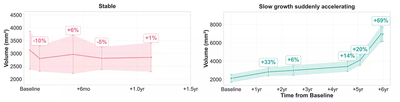
To meet the need for monitoring subtle but significant progression, the team developed a "one-click" method for longitudinal, quantitative tumor tracking that analyzes a patient's historical MRI data and provides a volume for each tumor at different time points. The algorithm runs immediately after the images are exported from the scanner, allowing the radiologist to see the algorithm’s comprehensive graph of tumor growth: stable, growing slowly, or progressing rapidly. This allows quick evaluation of treatment effectiveness, without waiting weeks or months for the next scan.
As Nedelec explained, “This gives a more holistic view of the evolution of the tumor, something that is hard to see today by just looking at individual exam MRIs.”
Path to Clinical Deployment
Nedelec and his collaborators are keen to ensure that users can trust the tool’s accuracy, a key step in clinical adoption. The system is designed to be transparent, allowing radiologists to easily see the AI's segmentation for any reported volume. Visiting graduate student Yassine Guennoun added confidence intervals to the tool, indicating the likelihood that a tumor’s reported volume is slightly larger or smaller than the actual size. UCSF’s AI Oversight Committee has approved the tool for internal research usage, and the team deployed the tool recently for a limited set of radiologists.
Nedelec said, “We’ll get feedback on what’s most useful and we’ll keep iterating. We hope to eventually deploy similar tools for other tumors as well.”
The Center for Intelligent Imaging (ci2) supported development of the tool’s front end. Now Nedelec and the developers are working to minimize bias within the algorithm by assessing the model's performance across different scanner types and patient demographics such as age and sex. The team is also adding a rating system to improve segmentation quality by incorporating physician feedback during testing.
Future directions for this tool show promise for monitoring other brain tumors, prostate cancer, or certain breast cancers. By training the algorithm on additional datasets, this tool could provide clinicians with precise, longitudinal data to help improve clinical decision making and overall patient care.