Musculoskeletal Magnetic Resonance Imaging Lab (Krug Lab)
The Musculoskeletal Magnetic Resonance Imaging Lab's main research interest relates to the assessment (MRI) and treatment (MR guided Focused UltraSound FUS) of lower back pain. Furthermore, we assess bone and bone marrow health, obesity and whole body composition, and osteoarthritis in various patient populations. In particular, we develop methods to assess:
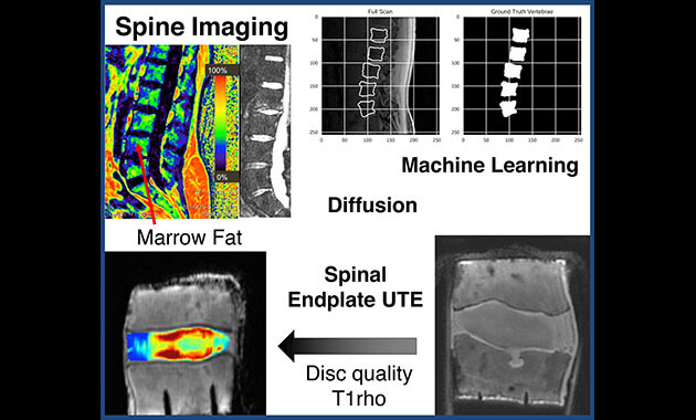
- Lower Back Pain (Biochemical Assessment of the Spinal Endplate using UTE, Intervertebral Disc T1rho, and Bone Marrow using Dixon).
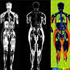
- Whole Body Composition (Visceral Adipose Tissue, Subcutaneous Adipose Tissue, Intramuscular Adipose Tissue, Muscle Volumes).
- Osteoporosis (Bone and Bone Marrow Quality in Hip, Wrist, Ankle, and Knee).
- Bone Hydration and Porosity.
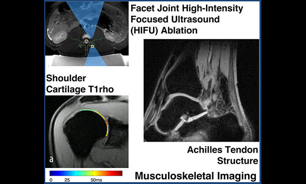
- High-Intensity Focused Ultra-sound (HIFU) for the Treatment of Lower Back Pain.
- Osteoarthritis (Assessment of the Calcified Cartilage Layer in the Knee).


News
Congratulations to Nico Sollmann for his work as a Trainee Deputy Editor of JMRI
June 2021
Journal of Magnetic Resonance Imaging (JMRI) Newsletter - June 2021 (pdf)

Research Published in Frontiers Endocrinology
The Krug Lab receives several NIH grants (>$30M) to study low back pain and opioid addiction
October 10, 2020
Featured it in UCSF News. Dr. Krug is the imaging director of the Core Center for Patient-centric Mechanistic Phenotyping in Chronic Low Back Pain (https://www.bacpac-reach.org/people-1) at UCSF (U19) as well as PI on a technogloy center (UH2/3).
 The Krug Lab's work is featured on the Cover of JMRI
October 2019 edition
Krug R et al. Associations between vertebral body fat fraction and intervertebral disc biochemical composition as assessed by quantitative MRI. JMRI 2019 Oct;50(4):1219-1226.
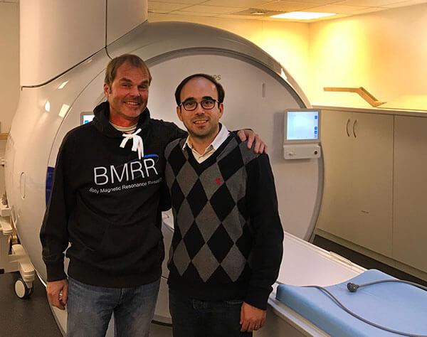
Body MR Imaging Group in Munich
September 2019
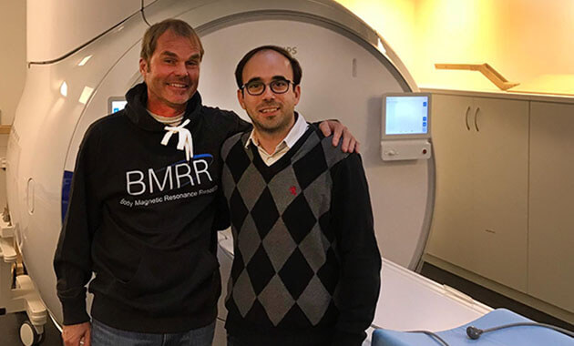
Visiting Professor at the Technical University of Munich. The Body MR Imaging group (BMRR) in Munich lead by Prof. Dimitrios Karampinos (image below right) is a strong collaborator of our laboratory.
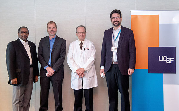
Celebration of 15 years at UCSF with Department Chair and Dean
Oral Presentations and Podium Talks
- ISMRM 2019 workshop on Obesity Singapore (Invited Talk)
- ISTU/EFUS 2019 in Barcelona. SI Joint pain ablation using MRgFUS. (Krug et al.)
- ORS 2018 in New Orleans. Quantitative MRI in patients with lower back pain (Krug et al.)
- ISMRM 2018 in Paris. Relationship of vertebral fat, and disc health in patients. (Krug et al.)
- ASBMR 2018 in Montreal, Canada. Bone Marrow Fat and its Associations with Bone Quality in the Proximal Femur. (Krug et al.)
- FUS Foundation 2018 in Virginia. MRgFUS for back pain. (Rieke, ..., Krug)
Featured on the Cover of QIMS (Feb 2018 edition)
G.J. Kazakia, J. Carballido-Gamio, A. Lai, L. Nardo, L. Facchetti, C. Pasco, C.A. Zhang, M. Han, A. Hutton Parrott, P. Tien, R. Krug. "Trabecular Bone Microstructure is Impaired in the Proximal Femur of HIV-Infected Men with Normal Bone Mineral Density"
Oral Presentation at the ORS 2018 in New Orleans
Krug et al.: Associations between Disc Biochemical Composition and Vertebral Body Fat Fraction using Quantitative MRI.
Research on Hip Osteoporosis featured on the title page of JMRI (May 2015 edition)
Han M, Chiba K, Banerjee S, Carballido-Gamio J, Krug R. "Variable flip angle three-dimensional fast spin-echo sequence combined with outer volume suppression for imaging trabecular bone structure of the proximal femur"
Research Projects
Quantifying Temperature Changes in Bone
We have recently published our research on temperature assessment of bone during heating. The goal of this research is to monitor temperature in bone in-vivo during high intensity focused ultrasound (HIFU) ablation.
High Resolution Assessment of Bone Marrow Fat Content
Our group has been pioneering the assessment of bone marrow fat content in the presence of trabecular bone with high spatial resolution. We have recently evaluated this technique in phantoms and compared in-vivo with MR spectroscopy:
- C. Gee, J. Nguyen, C. Marquez, J. Heunis, A. Lai, C. Wyatt, M. Han, G. Kazakia, A. Burghardt, D. Karampinos, J. Carballido-Gamio, R. Krug. Validation of Bone Marrow Fat Quantification in the presence of Trabecular Bone using MRI. JMagnResonImaging (2015 in press).
- D. Karampinos, G. Melkus, Th. Baum, J. Bauer, E.Rummeny and R.Krug. Bone marrow fat quantification in the presence of trabecular bone: Initial comparison between water-fat imaging and single-voxel MRS. Magn Reson Med. Volume 71, Issue 3, pages 1158–1165, March 2014.
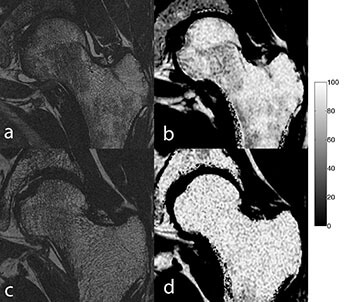
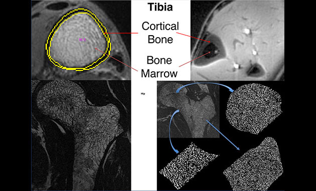
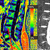
Figure shows typical in-vivo examples of high-resolution MRI images along with corresponding MRI Fat Fraction maps. The top row shows images from a young, healthy female. Image a) depicts the trabecular structure and image b) the corresponding Fat Fraction map. The high-resolution image shows a dense trabecular structure. The darker, hematopoietic bone marrow is clearly depicted and can be quantified on the Fat Fraction map b). The bottom row shows images from a postmenopausal woman who sustained a low impact fracture. The high-resolution image c) shows dense bone but the Fat Fraction map d) also reveals overall high bone marrow fat content as compared to b) where large areas of hematopoietic bone marrow are present.
Assessment of Temperature in Bone using MRI
We have recently shown for the first time that temperature changes can be measured in bone during heating using T1-UTE based MR imaging. We have presented these results at ISTU 2014 and ISMRM 2014 in oral presentations.
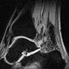
Bone Marrow Fat Fraction Mapping in the Proximal Femur in vivo using IDEAL
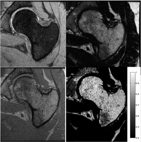
Figure: Clockwise from top left: Water only image, Fat only image, Fat Fraction Image and In-Phase image.
(D. Karampinos, R. Krug)
There is some evidence that osteoporosis is associated with increased marrow fat content as well as a conversion from red marrow to yellow (fatty) marrow with age. In this project, we investigate the marrow fat composition in the proximal femur in vivo using a method based on Iterative Decomposition of water and fat with Echo Asymmetry and Least-Squares Estimation (IDEAL). Three-point IDEAL FGRE hip images of six healthy subjects were acquired and water-fat separation was performed using multi-peak IDEAL. The average fat fraction and standard deviation were determined in three different regions of interest (femoral head, trochanter and neck). Significant differences in marrow fat content were identified between the three regions for all subjects.
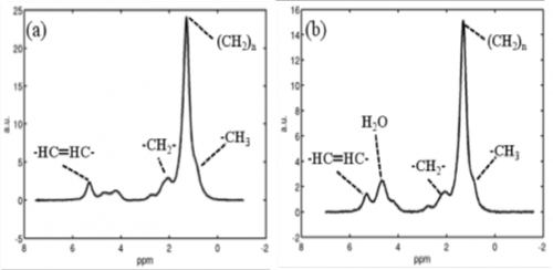 Figure 1: Comparison of single-voxel PRESS MR spectra from the trochanteric region (left) and the neck region (right). Note the enhanced water peak on the right spectrum.
Advanced Image Analysis Techniques of New High-resolution Images of the Proximal Femur in the Presence of Red and Yellow Bone Marrow using Local Bone Enhancement Fuzzy Clustering
(J. Folkesson, R. Krug)
Using advanced MR hardware and pulse sequences technique, we have been able to image the small trabecular structure of deeper sited seated regions like the proximal femur with very high spatial resolution in a clinically feasible scan time. We have developed and employed a novel partial membership bone segmentation technique (BE-FCM) that enhances bone segmentation compared to an established dual thresholding method in the presence of signal variations due to different marrow types. The new image acquisition and analysis framework enables trabecular bone analysis in the deeply situated femoral head, something which has been previously unfeasible in vivo.
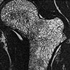
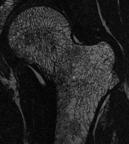 Figure 2: High-resolution in vivo image of the proximal femur. Hemopoietic or red marrow is composed of ~40% water (longer T1) and appears dark (femoral head, neck and shaft). The more prevalent yellow marrow is more fatty and yields a brighter MR signal.
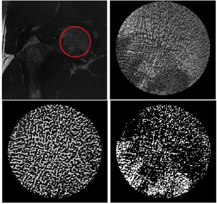 Figure 3: Top images: ROI defined in the femoral head. Bottom image (left): Trabecular bone segmentation of the ROI using the advanced BE-FCM technique. Bottom image (right): The same ROI segmented using an established dual thresholding technique.