Abdominal Imaging Division Research
 Abdominal imaging division research at UCSF is focused on developing and testing new methods for diagnosing diseases of the organs, systems and tissues of the abdomen and pelvis.
Clinical and preclinical research complements the clinical diagnostic efforts in the abdominal imaging subspecialty. Research questions are driven by clinical need and involve a multidisciplinary approach. Our abdominal imaging physicians actively publish papers on novel imaging methods and proper image interpretation—particularly for CT and MRI— and they routinely challenge myths regarding the significance of many commonly utilized diagnostic radiologic signs.
Collectively, our abdominal imaging physicians have published several hundred scientific papers, authored or co-authored multiple textbooks, and attracted more than $6 million in funding from the National Institutes of Health (NIH).
Current activities cover:
High Intensity Focused Ultrasound (HIFU) / MR Guided Focused Ultrasound Surgery (MRg-FUS)
High Intensity Focused Ultrasound (HIFU) (also known as MRg-FUS) at UCSF is an interdisciplinary effort that involves the active contribution of numerous interested investigators across multiple departments.
- Pre-procedural imaging and follow-up of uterine fibroids
- Prostate cancer ablation
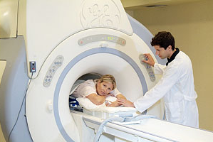
Patient on ExAblate Treatment Table.
